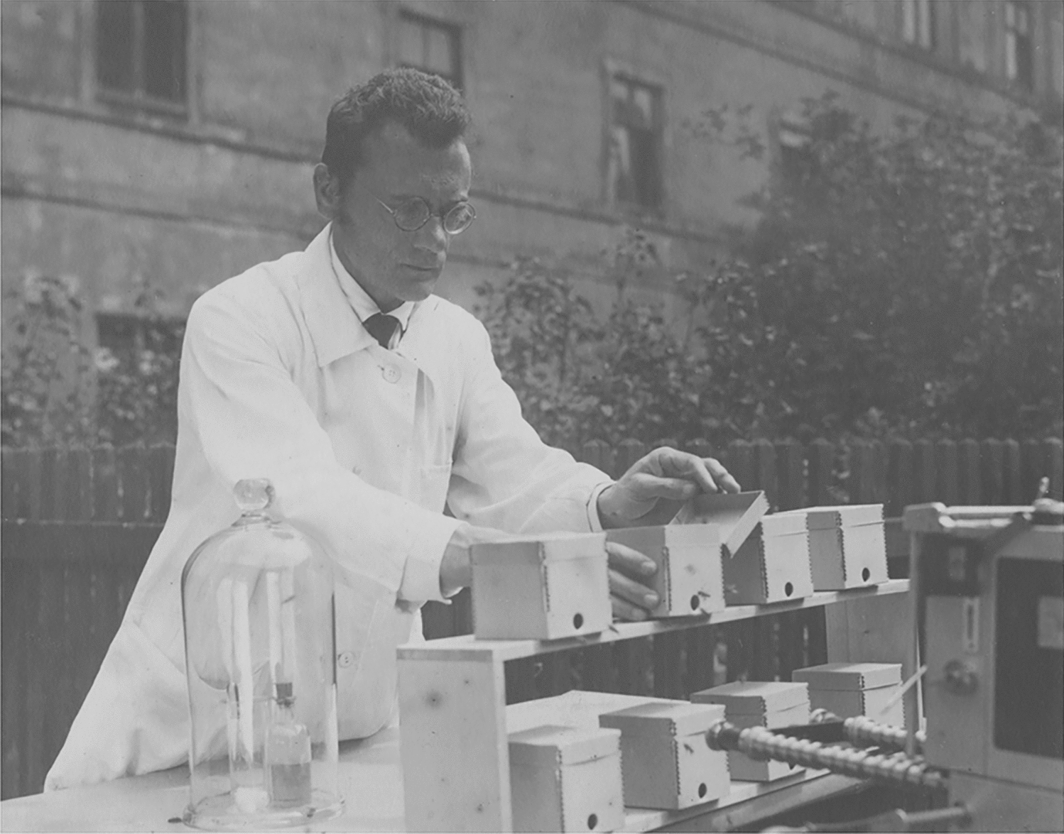
Artificial reproduction (AR)
To obtain F1-hybrids between male G. petersii and female C. compressirostris, we performed three independent AR experiments (Table 1), for which we selected males and females from the breeding tanks. We chose four different males, which could be identified based on their TL and their anal fin features. The females could not be identified individually, but were selected based on their body volume: thick females indicated well-developed ovaries; this was confirmed through the release of ovulated eggs in all three reproduction experiments (Table 1). Despite the different origin of the gametes in the three experiments, the fertilisation rates were very similar (20.1–25.3%) and the number of hatched embryos did not vary much (86.1–97.7%). However, the survival rates of the F1-hybrids varied considerably: between 0% and 48.4% (Table 1). The possible cause for this result will be discussed in the paragraph below. For comparison, the fertilisation rate of the interspecific hybridisation between male of G. petersii and female of C. rhynchophorus was 35% and the hatching rate amounted to 68.6% (Kirschbaum et al. 2016). The fertilisation rates of the eight intraspecific Campylomormyrus hybrids showed a wide range varying between 19.7% and 94% (average 47.8%) and the hatching rates ranged between 16.4% and 99% (average 82.1%) (Kirschbaum et al. 2016). Thus, fertilisation and hatching rates of the two intergenus hybrids (Gnathonemus × Campylomormyrus) were not consistently different from the respective values of the intragenus hybrids (Campylomormyrus).
Ontogenetic development and fertility
The F1-hybrids start to hatch on day 3 (about 70–72 h after fertilisation), exogenous feeding starts on day 11, the juvenile stage starts at a TL of ca. 21 mm (60–65 day-old-fish) and sexual maturity is achieved at a TL of 10–11 cm (Fig. 8). The embryos, the free embryos up to day 11 and the F1-hybrid larvae look like those of the parental species C. compressirostris (Nguyen et al. 2017) and G. petersii (Korniienko unpublished results). Furthermore, they are comparable to early ontogenetic stages of other mormyrid species (Kirschbaum 1987, 2006; Kirschbaum and Schugardt 2002a; Diedhiou et al. 2007; Nguyen et al. 2017), as species specific morphological features appear later in development. The transition from the larval to the juvenile stage occurs in the F1-hybrids at a TL of ca. 21 mm. This is similar to the development of C. compressirostris, C. rhynchophorus and C. tshokwe in which this transition occurs at a TL of about 20 mm (Nguyen et al. 2017). However, in smaller species this transition occurs at a smaller TL, e.g., in Pollimyrus isidori, which attains an adult TL of ca. 10 cm, it occurs at a TL of ca. 15–16 mm (Kirschbaum 1987). The minimum size for sexual maturity of the F1-hybrids was a TL of 10–11 cm (Fig. 8). C. compressirostris achieves maturity at a TL of 13–14 cm (Paul et al. 2015), in contrast to G. petersii, which attains sexual maturity at a TL of 15–16 cm (Korniienko unpublished data). Thus, the F1-hybrid G. petersii × C. compressirostris attains sexual maturity at a smaller size than the parental species. A similar phenomenon was observed in the F1-hybrid C. tamandua × C. compressirostris (Korniienko et al. 2020).
In our AR experiments with the F1-hybrids G. petersii × C. compressirostris, it was indeed shown that three females with a TL between 13.5 and 14.5 cm released a total of 1144 ovulated eggs, which proves that these females were fertile. From three males (sizes between 16 and 18.5 cm of a TL), used during this experiment, it was possible to obtain a liquid, which we interpreted as sperm; however, this liquid did not fertilise any egg in a batch of 366 eggs, which we selected from one of the three females. This was similar to the fertile F1-hybrids C. tamandua × C. compressirostris: not all the sexually mature males (identification based on the male-typical anal fin) chosen for the artificial breeding experiment gave sufficient sperm (Kirschbaum et al. 2016). The inter- and intragenus F1-hybrids, apparently, produce less sperm than the purebred species. To clarify this issue, histological investigations of the testis of the F1-hybrids would be helpful.
Despite a more transparent sperm liquid of the F1-hybrids, the fertile intragenus hybrids C. tamandua × C. compressirostris spawned naturally with an average fertilisation rate of 47.8% (Korniienko et al. 2020), which is quite high for mormyrid species (see e.g., Nguyen et al. 2017). As our first artificial breeding experiments with the intergenus hybrids G. petersii × C. compressirostris were not successful (see Results), it would be interesting to perform additional breeding experiments to find out, if they are also able to spawn naturally.
The fact that the intergenus F1-hybrids G. petersii × C. compressirostris are fertile indicates that the genetic difference between species of the two sister clades Campylomormyrus and Gnathonemus (Sullivan et al. 2000; Lavoué et al. 2003) are not large enough to prevent the occurrence of fertile F1-hybrids.
The morphology of the adult F1-hybrids G. petersii × C. compressirostris shows features of both parents, which is well seen in the morphology of the snout and the pigmentation of the trunk (Fig. 8). A more detailed analysis of this topic, based on landmarks and geometric morphometrics, is in progress (Amen unpublished results).
Malformations and mortality
Malformations (Figs. 3–5) were observed during embryogenesis, after hatching in the free embryos, during the larval stage and at the beginning of the juvenile stage. These included abnormalities of the vertebral column, disturbances in the circulatory system, abnormalities of the heart region and of the ventral part of the body cavity. All these deficiencies finally led to the death of the specimens (Fig. 6) indicating deficiencies in the genetic inventory of these F1-fish.
Malformations were also described in intragenus Campylomormyrus hybrids (Baumgartner 2015; Elarbani 2017). However, in these hybrids typically only one of the abnormalities occurred at a time, whereas in the intergenus hybrids often two or even more abnormalities were observed in individual animals. For instance, some of the F1-hybrids of C. tamandua × C. compressirostris showed eye, snout or trunk abnormalities, yet they developed otherwise normally (Baumgartner 2015). Also in the intergenus hybrids G. petersii × C. rhynchophorus, malformations occurred more frequently and included two or more defects, such as a concave neck region in combination with missing eyes and disorder in the yolk (Elarbani 2017). The higher degree of malformations in the intergenus hybrids is likely related to the genetic differences between the two clades (see Lavoué et al. 2003).
In our study, the F1-hybrids died within different periods of the ontogenetic development (Fig. 6) apparently caused by genetic deficiencies. Deaths at the beginning of exogenous feeding might indicate problems with the digestive system concerning food uptake; deaths at the transition from the larval to the juvenile period occurred, when we switched to larger food items and moved the fish into larger tanks.
The time course of deaths in our three breeding experiments differed considerably (Fig. 6). We observed in both breeding experiments I and II symptoms, which we interpreted, based on our experience with raising mormyrid fish, as indication of diseases. This led to a complete death of all the hybrid-fish in experiment II and to the death of all fish (except one) in breeding experiment I. Still, we do not know why these diseases did not or only to a small part affect the fish of breeding experiment III. Probably, some uncontrolled aspects of rearing (i.e., food intake, injuries through aggression) are contributing to these differences.
EOD
The larval EOD of the F1-hybrids is biphasic with about 3 ms duration (Fig. 9a). Such a larval EOD is produced by both parental species (Kirschbaum et al. 2016; Nguyen et al. 2017) and is found in other Campylomormyrus (Nguyen et al. 2017) and several other mormyrid as well (Westby and Kirschbaum 1977, 1978; Baier et al. 2006; Werneyer and Kramer 2006). At a TL of 20.5 mm, the EOD of the hybrids starts to change and at a TL of 24 mm, the larval EOD is replaced by the juvenile EOD (Fig. 9b–d). This biphasic juvenile EOD has a different shape than the larval EOD and is shorter in duration (duration of about 150–200 µs). It is similar to the juvenile EOD of the parental species (Kirschbaum et al. 2016; Nguyen et al. 2017; Korniienko unpublished results). At higher magnification, it becomes apparent that there are two distinct juvenile EOD types: a tetraphasic EOD type (Type I, Fig. 9e1) and a biphasic EOD type (Type II, Fig. 9e2). Type II EOD further develops into a triphasic EOD (Fig. 9f2, g2). Applying structural–functional correlations provided by Bass (1986) for mormyrid EOs, this suggests anatomical differences in the EO of the F1-hybrids. Specifically, the first small initial head negative phase of Type I EOD and the subsequent large head positive phase would be compatible with penetrating small stalks, which originate at the caudal surface of the electrocyte and a rostral position of the main stalk. In contrast, type II EODs is indicative of a caudal position of the main stalk and caudally located, non-penetrating small stalks. These results suggest that the morphological design of the EO in the paternal G. petersii (rostral position of the main stalk and penetrating small stalks; Bruns 1971) has been transferred to the F1-hybrids. This was not the case in the hybrids of the cross G. petersii × C. rhynchophorus (Kirschbaum et al. 2016; Elarbani 2017). The split into two EOD types (Fig. 9e1–g1, e2–g2) in our sample indicates that this feature of the G. petersii EO is only inherited by ca. 65% of the hybrids (see Fig. 8). Heterozygous genetic background of the parental species, controlling the morphology of the EO, might be responsible for this divergence. Another explanation could be that, during early ontogeny, the structure of the hybrid EO is not yet fixed and underlies epigenetic influences in the one or the other direction.
Several recent studies in Campylomormyrus hybrids (Kirschbaum et al. 2016) and in wild populations of Paramormyrus (Gallant et al. 2011; Picq et al. 2020) argue for a further diversity in EO design in specimens with EODs containing an initial head negative (pre-potential) similar to Type I:
(1)
Hybrids of C. tamandua and C. compressirostris feature electrocytes with double penetrations but caudally located stalks (Kirschbaum et al. 2016). This is comparable to the EO in Stomatorhinus corneti (Bass 1986).
(2)
Hybrids of C. tamandua and C. tshokwe feature an EO of mixed morphology, where inside the column of electrocytes some cells possess a rostral and some a caudal position of the main stalk both featuring penetrations (Kirschbaum et al. 2016). Heterogeneously organised EOs were so far only reported in some populations of Paramormyrops kingsleyae from Gabon (Gallant et al. 2011; Picq et al. 2020).
(3)
In Paramormyrops kingsleyae, there is significant geographic variation in electric signal waveforms with some specimens exhibiting initial head negative (pre-potentials)–similar to Type I EODs in our hybrids–and others lacking them completely–similar to our Type II EODs. In Paramormyrops, the magnitude of the first head negative phase of the EOD (called “P0”) positively correlates with the number of penetrations per area of electrocytes (Gallant et al. 2011).
Further studies are needed to investigate the morphological basis of EOD diversity in the intergenus F1-hybrids. Picq et al. (2020) have demonstrated that Paramormyrops kingsleyae is capable of distinguishing between EODs with pre-potential and without. This suggests that this kinds of EOD variation could be a cue for assortative mating (Picq et al. 2020). Such a mixture of two EOD types is also found in our intergenus F1-hybrids. The breeding experiment to obtain natural spawnings with the F1-hybrids failed. Possibly, because the individuals with identical EODs could not segregate well enough in the breeding group in the restrictive space of the breeding conditions. Further behavioural experiments are necessary to assess this issue.
Species stability in mormyrid fish
Hybrids are commonly observed in fishes and occur in more than 19.7% of the fish families worldwide (Nelson 1994). In marine species, hybrids are widely found in the families belonging to the Atherinomorpha and Percomorpha (Schwartz 1972, 2001). Hybrids in freshwater fish have been documented in 30 families (Schwartz 1981, 2001). Molecular genetic studies have repeatedly shown that hybridisation in fish is a common phenomenon and is often detected as an ancient introgression (Schliewen and Klee 2004; Herder et al. 2006; Schwarzer et al. 2012a, 2012b; Meier et al. 2017; MacGuigan and Near 2019). In the freshwater family of the Mormyridae with more than 200 species (Lavoué et al. 2003), natural hybrids are only rarely observed. Natural hybridisation has been found only in the genus Paramormyrops, which contains 22 species (Lavoué et al. 2008): hybrids were observed between morphs of the Paramormyrops magnostipes species complex characterised by differences of their electric organ discharge (Arnegard et al. 2005). In Paramormyrops kingsleyae, natural hybrids were observed among morphs occurring in geographic proximity (Gallant et al. 2011). Sullivan et al. (2004) discuss past introgression (i.e., hybridisation) in Paramormyrops inferred from mitochondrial data. Within Campylomormyrus, mitochondrial and single locus nuclear data do not reveal any sign of hybridisation/introgression (Feulner et al. 2007; Lamanna et al. 2016), while recent genome-wide Single-Nucleotide Polymorphism (SNP) data point towards the possibility of one or two ancient introgression events among species of this genus (Canitz, Kirschbaum and Tiedemann unpublished data).
The AR experiments in the mormyrid fish (Kirschbaum et al. 2016; Korniienko et al. 2020; this paper) have documented a high potential in mormyrid fish to generate fertile hybrids, such that postzygotic isolation seems weak or even absent. In contrast, natural hybrids seem to be rare (see above), pointing towards effective prezygotic isolation. Indeed, the EODs are very diverse and serve as reproduction isolation barriers, as association with conspecifics is strongly preferred (e.g., Feulner et al. 2009a; Gallant et al. 2011; Nagel et al. 2018; Picq et al. 2020). Such close contact is essential during spawning, as the male’s anal fin forms a pouch into which eggs and sperm are released (Crawford et al. 1986; Kirschbaum 1987). As the mormyrid sperm is lacking a flagellum (Mattei et al. 1972; Pecio 2020), free sperm are rarely available for fertilisation of eggs of other mormyrid species. EOD-based mate choice and the peculiarities of sperm and the fertilisation process apparently act in conjunction as very effective prezygotic isolation mechanisms leading to the high species stability of the mormyrid fish, despite of an apparent lack of postzygotic isolation.


留言 (0)