
記住我
During central nervous system (CNS) development, neural circuitry formation involves complex interactions of molecular guidance and recognition cues. Members of the Teneurin protein family function as such cues and promote cell-cell adhesion through homophilic and heterophilic interactions (Rubin et al, 2002; Silva et al, 2011; Hong et al, 2012; Mosca et al, 2012; Beckmann et al, 2013; Boucard et al, 2014; Berns et al, 2018; Sando et al, 2019; Pederick et al, 2021). Teneurin proteins are large evolutionarily conserved dimeric type II transmembrane proteins, with an intracellular domain that is responsible for downstream signaling (Rubin et al, 2002; Tucker et al, 2012). Early works in Drosophila have shown that Ten-a and Ten-m function as homophilic matching cues in the olfactory bulb and in the neuromuscular junction (Hong et al, 2012; Mosca et al, 2012). In mammals, Teneurin has four paralogs that are predominantly expressed in the brain, with partially overlapping expression patterns from early embryonic development to adulthood (Ben-Zur et al, 2000; Zhou et al, 2003). Teneurins have been shown to play crucial roles in the development of retinal, hippocampal, and cortical circuits. In fact, recent mouse genetic experiments have revealed that the concomitant axonal and dendritic expression of Teneurin3 promotes the correct neuronal pathfinding and wiring in the mouse hippocampus (Berns et al, 2018; Pederick et al, 2021).
Besides homophilic interactions, Teneurin proteins can also establish heterophilic interactions with a member of the adhesion G-protein-coupled receptors, known as Latrophilin (Silva et al, 2011; Boucard et al, 2014; Vysokov et al, 2016; Li et al, 2020; Pederick et al, 2021). Latrophilins themselves have been shown to interact with fibronectin leucine-rich repeat transmembrane proteins (FLRTs; O'Sullivan et al, 2012; Lu et al, 2015; Ranaivoson et al, 2015; Jackson et al, 2016). Coincident interaction of Latrophilin with its two binding partners (Teneurin and FLRT) potentiates the formation of excitatory synapses in the mouse hippocampus (Silva et al, 2011; Boucard et al, 2012; Sando et al, 2019). Notably, interfering with the Latrophilin–Teneurin interaction specifically has resulted in a decrease in excitatory synapse formation (Li et al, 2020). More recently, Del Toro et al. have established an additional role for Teneurin2 in complex with Latrophilin and FLRT in cortical cell migration in vitro and in vivo (Del Toro et al, 2020).
In humans, Teneurins have been associated with specific neuronal disorders, such as essential tremor, microphthalmia, general anosmia, schizophrenia, and bipolar disorder (Burbach & Meijer, 2019), highlighting the biomedical need to better understand how Teneurins function and orchestrate the formation of neuronal networks.
In the recent years, structural characterizations of the partial ectodomain of Teneurin2 and Teneurin3 by crystallography and cryo-electron microscopy have revealed an extracellular ~1,900 residue superfold, with no resemblance to other typical cell-adhesion proteins (Jackson et al, 2018; Li et al, 2018). The C-terminal region of the ectodomain is folded into a large barrel-shaped structure, termed YD-shell, with a beta-propeller NHL domain positioned at an almost 90° angle. The barrel is sealed upstream by a so-called fibronectin plug domain and capped downstream by its own inward spiraling C-terminal. The YD shell, together with the NHL domain, resembles a bacterial toxin system known as TcB and TcC of Y. entomophaga and P. luminescens, now known to be present in other bacterial strains as well (Jackson et al, 2018, 2019). Two additional domains, the antibiotic-binding like (ABD) and Tox-GHH domains, are located at the ultimate C-terminal region and appended onto the YD shell. Finally, the transthyretin (TTR)-like domain is the most N-terminal domain of the superfold and wedges in between the FN-plug and NHL domain. Eight predicted epidermal growth factor (EGF)-like domains are located upstream of the Teneurin superfold. EGF2 and EGF5 each harbor a free cysteine that enable covalent cis-dimerization. Interaction of a confined region on the YD-shell, opposite of the ABD and Tox-GHH domains, with Latrophilin, possibly in trans (opposite cells), might form the basis of a ternary complex consisting of Teneurin, Latrophilin, and FLRT connecting the pre- and postsynaptic membrane (Del Toro et al, 2020; Li et al, 2020). Interestingly, Teneurin-Latrophilin binding is prohibited by an NHL splice insert version of membrane-bound Teneurin (Boucard et al, 2014; Li et al, 2020).
So far, detailed structural characterizations dealt with monomeric versions of Teneurin proteins (Jackson et al, 2018; Li et al, 2018, 2020; Del Toro et al, 2020). However in biological systems, Teneurins are expressed on the cell surface as covalently bound dimers by means of two disulfide bonds in the extracellular EGF-like repeats 2 and 5 (Feng et al, 2002). How would macromolecular complex assembly further be supported by the homodimeric conformation of Teneurins? Using cryo-electron microscopy, X-ray diffraction (XRD), small angle X-ray scattering (SAXS), and thermostability assays, supported by cell biological assays, we show here that dimeric Teneurin4 can adopt a compact architecture for complex assembly allowing both cis- and trans-interactions. We identify three calcium-binding sites in the previously unreported C-rich domain and show that calcium stabilizes the compact Teneurin4 ectodomain conformation. The dimeric nature of Teneurin might be instrumental for the clustering of cell adhesion complexes to establish functional circuits in the developing brain.
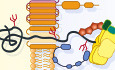
Results Covalent dimers of human Teneurin4 are compact moleculesTo determine the structure of covalently linked Teneurin dimers, we expressed the complete ectodomain of human Teneurin4 in HEK293 cells (Fig 1A). The protein was purified using affinity purification followed by size-exclusion chromatography (Fig EV1A) and a 3.5 Å structure of the covalent homodimer was determined with single-particle cryo-electron microscopy (Figs 1B, EV1B–J, and Table 1), with the core, that is, FN-plug, YD-shell, linker, ABD, and Tox-GHH combination, resolved to 3.2 Å (Fig EV1G and J). The structure revealed a novel compact conformation of the dimeric molecule. In order to obtain high-resolution information, we inserted a stabilizing disulfide bond between the two ABD domains (S2585C, Teneurin4Mut) located in the core of the homodimer (Fig 1A and C inset).

Figure 1. Human Teneurin4 ectodomain adopts a compact-dimer structure
Domain composition, including the covalently dimerizing cysteines in EGF repeats 2 and 5 (C597 and C696). The S2585C mutation (Teneurin4Mut) was introduced to stabilize the compact dimer conformation (in green). Predicted glycans are indicated as thunderbolts. Regions in grey are not represented in the cryo-EM structure. ICD, intracellular domain; Ig, immunoglobulin fold; EGF, epidermal growth factor repeat domain; C-rich, cysteine-rich region; TTR, transthyretin-related; FN-plug, fibronectin plug; NHL, NCL, HT2A and Lin-41; YD, tyrosine-aspartate; ABD, antibiotic-binding domain; Tox-GHH, toxin-glycine-histidine-histidine. Overlay of cartoon representations of human Teneurin4WT (purple) and human Teneurin4Mut (grey) with one chain in transparent surface to indicate the two dimer subunits. The S2585C mutation does not influence the structure of the compact Teneurin4 dimer. N and C indicate the N- and C-termini, respectively. Cartoon representations of human Teneurin4Mut colored by domain as indicated in (A) with the S2585C mutation, introducing an intermolecular disulfide bond, as indicated in the inset. Scale bar, 20 Å. Click here to expand this figure.
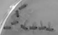
Figure EV1. Related to Fig 1. Image reconstruction of Teneurin4WT
Size-exclusion chromatography traces of purified Teneurin4WT dimer (blue) and Teneurin4Mut dimer (red). The difference in peak retention volume may indicate a size difference of the two Teneurin4 samples, with a smaller (or more compact) size for the Teneurin4Mut dimer. Inset shows Coomassie-blue-stained SDS-Page gel in presence and absence of β-mercaptoethanol (BME) and confirms both samples are covalent dimers. Representative electron micrograph of Teneurin4WT. Scale bar, 40 nm. Selected 2D classes of Teneurin4WT. Scale bar, 10 nm. Workflow for Teneurin4 reconstruction. Note that a small subset of particles represents compact dimers, and a large subset represents extended dimers that have different conformations resulting in the reconstruction of a single subunit only. C1 symmetry reconstruction of Teneurin4WT compact dimer colored by local resolution as in (G). C2 symmetry reconstruction of Teneurin4WT compact dimer colored by local resolution as in (G). Reconstruction of Teneurin4WT core colored according to local resolution. Same as (E), but rotated by 180° over the x-axis. Structure of Teneurin4WT in cartoon representation with EGF repeat 6–8 depicted as density maps. Fourier Shell Correlation of structures shown in E–G. Dotted line represents FSC = 0.143. Table 1. Cryo-EM data collection and refinement statistics.Ten4WT
core focused
Ten4WT
C1
Ten4WT
C2
Ten4Mut
C2
Ten4Mut
single subunit
Ten4Mut
TTR – C-rich focussed
EMD-12122 EMD-12123 EMD-12124 EMD-12125 EMD-12126 EMD-12127 7BAM 7BAN 7BAO Data collection and processing Magnification 75,000× 75,000× 75,000× 165,000× 165,000× 165,000× Voltage (kV) 300 300 300 300 300 300 Electron exposure e-/Å2 50 50 50 50.6 50.6 50.6 Defocus range (µm) −2.8 to −1.1 −2.8 to −1.1 −2.8 to −1.1 −1.75 to −0.75 −1.75 to −0.75 −1.75 to −0.75 Pixel size (Å) 0.878 0.878 0.878 0.842 0.842 0.842 Symmetry imposed C1 C1 C2 C2 C1 C1 Initial particle images (n) 564,393 564,393 564,393 609,582 609,582 609,582 Final particle images (n) 27,751 35,929 35,929 242,300 212,571 188,064 Map resolution (Å) 3.2 3.7 3.5 2.7 2.7 3.4 FSC threshold 0.143 0.143 0.143 0.143 0.143 0.143 Map resolution range (Å) 3.2–4.6 3.6–7.0 3.4–6.2 2.6–4.0 2.7–3.7 3.2–4.4 Refinement Initial model used (PDB code) 6FB3 6FB3 6FB3 Model resolution (Å) 3.5 2.8 2.8 FSC threshold 0.5 0.5 0.5 Model composition Non-hydrogen atoms 30612 30612 15306 Protein residues 3808 3808 1904 Ligands (glycans & Ca2+) 36 36 18 B factors (Å2) Protein 73.1 66.4 64.8 Ligand 87.6 70.9 73.0 R.m.s. deviations Bond lengths (Å) 0.007 0.004 0.011 Bond angles (°) 0.77 0.78 0.982 Validation MolProbity score 2.26 1.82 1.83 Clash score 24.0 7.9 8.0 Poor rotamers (%) 0.36 0.18 0.18 Ramachandran plot Favored (%) 94.2 94.2 94.1 Allowed (%) 5.8 5.8 5.9 Disallowed (%) 0 0 0This provided a 2.7 Å reconstruction of the compact dimer conformation (Figs 1B and C, and EV2A–G). Alignment of the dimeric Teneurin4WT and Teneurin4Mut structures indicates that the structure of Teneurin4Mut is virtually identical to the Teneurin4WT protein (root mean square deviation (r.m.s.d.) = 0.5 Å for all 3,808 Cα atoms, Fig 1B). Dimensions of the compact dimer are 166 Å by 133 Å by 112 Å. A total of 12 predicted glycans on the extracellular domain (ECD) are expected to expand these dimensions (Figs 1A and EV5C); however, they are not fully resolved in our cryo-EM maps due to their flexible nature. A two-fold symmetry axis of the structure is positioned between the loops of the ABD domains (I2584–N2588) and the beta-strands of the YD-shells (L1660–S1686). The C1 reconstruction of the Teneurin4WT dimer reveals that EGF repeats 6–8 are positioned close to the YD shell and ABD domains, and that EGF8 links to a previously unresolved domain containing a cysteine-rich region that we denote C-rich domain (Fig EV1H and I). The ECD segment preceding EGF6 is not resolved in the reconstructed map and EGF repeats 6–8 are of considerably lower resolution (> 5 Å) compared to the rest of the ECD region (Fig EV1H). It is thus unclear if the EGF repeats have direct contacts with the YD shell and ABD domains in the dimer. Conceivably, the EGF repeat domain as a whole, that harbors the conserved intermolecular disulfide bonds in EGF2 and EGF5, contributes to stabilizing the compact dimer composition.
Click here to expand this figure.
Figure EV2. Related to Fig 1. Image reconstruction of Teneurin4Mut
Representative electron micrograph of Teneurin4Mut. Scale bar, 40 nm. Selected 2D classes of Teneurin4Mut. Scale bar, 10 nm. Workflow for Teneurin4Mut reconstruction. C2 symmetry reconstruction of Teneurin4Mut colored by local resolution. Coloring as in (E). Symmetry expanded half-dimer reconstruction of Teneurin4Mut colored by local resolution. The map quality of the C-rich region, TTR, FN-plug and NHL domains is improved in this reconstruction. Three different side views of a focused reconstruction of Teneurin4Mut C-rich–TTR. Top panel same orientation as D and E. Fourier Shell Correlation of structures shown in D–F. Dotted line represents FSC = 0.143.The compact dimer interface is formed by interactions of the ABD domain with the YD-shell and C-rich region of the other monomer (Fig 2A and B). Specifically, residues R2593, E2589, and R2639 of the ABD domain interact with two loops of the YD shell (T1636 in the loop spanning residues 1634–1638 and M1654-G1655-T1656 in the adjacent loop spanning residue 1,654–1,659, Fig 2Cpanel II and III), while R2662 of the ABD domain interacts with Q880 of the C-rich region (Fig 2C panels I and IV). The 2472 Å2 buried surface area in the dimer interface is predominantly hydrophilic with only very few hydrophobic contacts. The ABD domain is central in the interface and contributes most to the buried surface area (Table 2, Fig 2D). Notably, the potential furin cleavage site RTRR (Vysokov et al, 2016) (amino acids 2,659–2,662 in human Teneurin4) in the ABD domain is intact and partially buried in the dimer interface, precluding enzymatic processing in this compact conformation.

Figure 2. Dimer interface of human Teneurin4
Cartoon representation of the Ten4Mut dimer, with one subunit also shown as transparent surface model. Dashed rectangle indicates area shown in (C) (central panel). Domains are colored as in Fig 1A. Open book representation and surface model of domains involved in the dimer interface. Residues involved in the dimer interface are shown as spheres. Colors of the domains are shown as in Fig 1A. The ABD domain (green) contacts the YD-shell (blue) and the C-rich region (pink). Central panel depicts overview of the dimer interface. Roman numbers in mid-panels refer to location in central panel. Outer panels are rotated and zoom ins for better visualization of interacting residues. Hydrogen bonds are indicated by yellow dashed lines. Sequence alignment of all four human Teneurins. Residues that are part of the dimer interface are highlighted in the color of the interacting domain of the other monomer, and # represents the residues buried in the dimer interface by a surface area higher than 5 Å2. Table 2. Domain-specific buried surface area (BSA) in Å2. Total BSA (Ų) C-rich 234 TTR 0 FN-plug 0 NHL 0 YD shell 378 Linker 0 ABD 623 Tox-GHH 0A structural comparison with all three published Teneurin ECD monomer subunits (Jackson et al, 2018; Li et al, 2018) reveals a striking resemblance (Fig 3A), with r.m.s.d. scores ranging from 1.8 Å to 2.0 Å. Specifically, the r.m.s.d. score for hTen2 vs hTen4 is 2.0 Å for 1,230 Cα atoms; for chTen2 vs hTen4, 1.8 Å for 1,735 Cα atoms; and for mTen3 vs hTen4, 1.9 Å for 1,431 Cα atoms. This similarity is also observed at the single domain level (Fig EV3C–F), exposing only two minor structural differences. First of all, we observed an extra disulfide bond between C1035 of the FN-plug and C2524 of the linker domain, covalently locking the core domains into the so-called superfold (Fig 3B). Based on sequence analysis, this disulfide bond is also present in human Teneurin1, but not in human Teneurin2 or human Teneurin3 (Fig 3C). Furthermore, the arrangement of two beta-strands (N2646–Y2670) in the ABD domain of Teneurin4 is deviating from the other Teneurins (Fig EV3F). This could be explained by the compact dimer arrangement of Teneurin4, where this specific loop is part of the dimer interface, while the structures of all other Teneurins were determined from monomerized samples. The overall high structural similarity of the family members, in combination with the nonoverlapping expression patterns as described previously (Ben-Zur et al, 2000; Zhou et al, 2003), suggests a high level of functional redundancy. Indeed, in vitro binding studies of Teneurins and Latrophilins—where context plays no role—reveal substantial promiscuity (Burbach & Meijer, 2019). Altogether, our cryo-EM density maps reveal a novel compact conformation of covalently dimerized Teneurin4, while maintaining the overall structure of the superfolds.

Figure 3. Structures of Teneurins are similar
Overlay of cartoon representations of human Teneurin2 (6CMX) (Li et al, 2018), chick Teneurin2 (6FB3) (Jackson et al, 2018), mouse Teneurin3 (6FAY) (Jackson et al, 2018) and human Teneurin4 (7BAM; this paper). R.m.s.d 1.8 Å (over 1735 Cα atoms) for chick Teneurin 2 and human Teneurin4, 2.0 Å (over 1230 Cα atoms) for human Teneurin2 and human Teneurin4 and 1.9 Å (over 1431 Cα atoms) for mouse Teneurin3 and human Teneurin4. Cartoon representation of the intramolecular disulfide bond between C1035 of the FN-plug domain (red) and C2524 of the linker region (yellow) in human Teneurin4. Sequence alignment of the FN-plug—linker cysteines and flanking residues. The interdomain disulfide-forming cysteines are in bold and indicated by a bracket. The consensus symbols are according to Clustal Omega. Click here to expand this figure.
Figure EV3. Related to Figs 2 and 3. Domain-specific comparisons of published Teneurin structures
Surface representation of evolutionary conserved score (left panel) and dimer interface residues (right panel), colored as in Fig 1A. Amino acid alignment of interface residues in vertebrates. Coloring as in Fig 2D. Overlay of cartoon representations of the TTR domain of chick Teneurin2 (6FB3; green) and human Teneurin4 (7BAM; blue), r.m.s.d. 1.0 Å for 80 Cα atoms. Overlay of cartoon representations of the FN-plug domain of chick Teneurin2 (6FB3; green), mouse Teneurin3 (6FAY; orange) and human Teneurin4 (7BAM; blue), r.m.s.d. 0.8 Å over 185 Cα atoms for chick Teneurin2 and human Teneurin4, and 1.4 Å over 185 Cα atoms for mouse Teneurin3 and human Teneurin 4. Overlay of cartoon representations of the NHL domain of chick Teneurin2 (6FB3; green), mouse Teneurin3 (6FAY; orange) and human Teneurin4 (7BAM; blue). Highest r.m.s.d. 1.2 Å over 326 Cα atoms for mouse Teneurin3 and human Teneurin 4. Overlay of cartoon representations of the ABD and Tox-GHH domains of human Teneurin2 (6CMX; pink), chick Teneurin2 (6FB3; green) and human Teneurin4 (blue). R.m.s.d. of both domains equals 1.0 Å over 135 Cα atoms for human Teneurin2 and human Teneurin4, and 0.8 Å over 189 Cα atoms for chick Teneurin2 and human Teneurin4. Cysteine-rich domain structure reveals three calcium-binding sitesUsing a combination of XRD and cryo-EM, we determined the structure of the previously unresolved EGF8—TTR domain-linking segment (residues 834–919), that includes the highly conserved C-rich region (Figs 4A and EV4I; Tucker et al, 2012). The cryo-EM map reveals a compact domain (residues 834–871, denoted C-rich domain) (Fig EV2C, F and G) at the N-terminal side of this linker that contains eight acid amino acids and six cysteines. We determined a 1.5 Å resolution crystal structure of this Teneurin4 C-rich domain (Fig 4B–E, Table 3). In the C-rich domain, three calcium ions are fully coordinated, each in octahedral geometry, by side chains of the eight conserved acidic residues (E835, D840, D843, D845, D847, D851, D854, and D856), as well as N844, the backbone carbonyl oxygens of A837, K842, and L849, and two water molecules (Figs 4E and EV4A and B). The coordination of two calcium ions (I and II in Fig 4D and E) is similar to Thrombospondin type 3 repeats (T3) with a matching calcium-binding motif (Kvansakul et al, 2004; Figs 4A and EV4C) and the coordination of the third calcium resembles that of LDL receptor type-A (LA) modules (Blacklow, 2007; Yasui et al, 2010; Fig EV4D). The six cysteines in the C-rich domain form three disulfide bonds in the pattern 1–3, 2–5, 4–6 (C838–C857, C852–C863, and C858–C869, Fig 4D) also observed in LA modules (Yasui et al, 2010). The C-terminal side of the C-rich domain (residues 857–869) is stru
留言 (0)