
記住我


Breast cancers that express hormonal receptors (HR) and HER2 display resistance to targeted therapy. Tumor-promotional signaling from the HER2 and estrogen receptor (ER) pathways converges at the cyclin D1 and cyclin-dependent kinases (CDK) 4 and 6 complex, which drives cell-cycle progression and development of therapeutic resistance. Therefore, we hypothesized that co-targeting of ER, HER2, and CDK4/6 may result in improved tumoricidal activity and suppress drug-resistant subclones that arise on therapy. We tested the activity of the triple targeted combination therapy with tucatinib (HER2 small-molecule inhibitor), palbociclib (CKD4/6 inhibitor), and fulvestrant (selective ER degrader) in HR+/HER2+ human breast tumor cell lines and xenograft models. In addition, we evaluated whether triple targeted combination prevents growth of tucatinib or palbociclib-resistant subclones in vitro and in vivo. Triple targeted combination significantly reduced HR+/HER2+ tumor cell viability, clonogenic survival, and in vivo growth. Moreover, survival of HR+/HER2+ cells that were resistant to the third drug in the regimen was reduced by the other two drugs in combination. We propose that a targeted triple combination approach will be clinically effective in the treatment of otherwise drug-resistant tumors, inducing robust responses in patients.
IntroductionBreast cancers that express hormone receptors (HR) and have amplified human epidermal growth factor receptor 2 (HER2) are an aggressive subtype associated with visceral and brain metastases and increased resistance to targeted therapies (1–3). More than 30,000 women are diagnosed with HR+/HER2+ breast cancer every year in the United States.
Multiple endocrine therapies are available to block estrogen receptor (ER) signaling in patients with HR+/HER2+ disease. These include the selective ER modulator tamoxifen, the ER degrader fulvestrant, and aromatase inhibitors (e.g., anastrozole, letrozole, and exemestane). HER2-targeting agents, including the front-line monoclonal antibodies trastuzumab and pertuzumab, are also given to patients with HR+/HER2+ disease. However, HR+/HER2+ tumors are often resistant to dual ER and HER2-targeted therapies, which produced promising results in preclinical models (4), but yielded less impressive results in the clinical setting. In two randomized phase III clinical trials of combination targeted therapy, blocking ER and HER2 signaling in patients with HR+/HER2+ metastatic breast cancer showed only modest improvement of progression-free survival (PFS) and no improvement of overall survival (OS; refs. 5, 6). Resistance to dual targeted combinations has been attributed to a cross-talk between ER and HER2 and their downstream effectors (7–9). As such, until more efficacious targeted combinations are developed, patients with HR+/HER2+ metastatic disease continue to receive chemotherapy-containing regimens associated with multiple negative side effects and decreased quality of life (10). To this effect, recent advances in the development of novel HER2-targeted agents led to clinical approval of tucatinib, a potent and highly selective inhibitor of HER2 tyrosine kinase activity (11). In addition to its beneficial oral availability, tucatinib has demonstrated impressive efficacy in combination with chemotherapy and other HER2-targeted agents in heavily pretreated patients with metastatic HER2+ breast cancer (12–14). Unlike most HER2-targeting antibodies, which are unable to cross the blood–brain barrier, tucatinib is able to penetrate in the central nervous system and treat brain metastasis (13, 15).
Progression of cancer cells through the cell cycle is regulated by cyclins and cyclin-dependent kinases (CDK). Signaling cross-talk between the HER2 and ER pathways converges at cell-cycle checkpoints where HER2 and ER both increase transcription of CCND1, the gene encoding cyclin D1 (16). Upon a mitogenic stimulus, cyclin D1 complexes with CDKs 4 and 6 to phosphorylate retinoblastoma (RB) protein, inducing cell-cycle progression. The cyclin D1–CDK4/6 complex has been proposed as a key molecular driver of tumorigenesis via ER and HER2-mediated signaling and a primary driver of therapeutic resistance to drugs targeting the ER and HER2 pathways (17–19). Indeed, cyclin D1 expression is a prerequisite for development of the tumors driven by HER2, as cyclin D1-deficient mice do not develop mammary tumors in HER2-driven models (20). Furthermore, the cyclin D1–CKD4/6 complex also controls ER-dependent cell-cycle progression; moreover, cyclin D1 binds to ER and increases its transcriptional activity, which creates a positive feedback loop (21). The CDK4/6 inhibitor palbociclib is FDA approved for treatment of patients with HR+/HER2-negative metastatic breast cancer based on the PALOMA clinical trials, where it showed marked improvement in median PFS in combination with endocrine therapies, including fulvestrant (22, 23). Recently, evidence for synergy between CDK4/6 inhibitors with both ER and HER2-targeting agents was shown in preclinical studies (17, 19, 24). Yet, insufficient research exists on the use of CDK4/6 inhibitors in HR+/HER2+ breast cancer, and no CDK4/6 inhibitors have been approved in combination therapy for HER2+ disease.
We propose that a novel triple targeted combination of tucatinib, fulvestrant, and palbociclib could overcome resistance to ER and HER2 inhibition driven by cyclin D1-CDK4/6 in HR+/HER2+ disease. We show that this combination in HR+/HER2+ human breast cancer cell lines and cell line–based xenograft models reduces viability in vitro and growth in vivo. In addition, we show that drug resistance to a single agent in triple combination is abrogated by the other two agents. Finally, we report that the triple combination works against a specific drug resistance mechanism that is present in 35% of all HER2+ breast tumors—overexpression of cyclin E (25, 26).
Materials and MethodsCell lines, media, and reagentsBT474 (RRID:CVCL_0179), MDA-MB-361 (RRID:CVCL_0620), and UACC812 (RRID:CVCL_1781) cells were obtained from the ATCC and cultured in standard conditions. Trastuzumab-resistant BT474-HR20 cells were kindly provided by Dr. Bolin Liu (Louisiana State University School of Medicine, New Orleans) (27). Cells were Mycoplasma free (last test October 9, 2020) and STR-profiled for all studies. Non-resistant cells were used under passage 10. Drug-resistant subclones of BT474 and MDA-MB-361 were generated by culturing cells with step-wise increasing concentrations of inhibitors over 12 months (28). Tucatinib was provided by Seattle Genetics. Palbociclib and fulvestrant were purchased from Selleckchem.
Cell viability assaysCell viability was measured by Cell Titer Glo assay (Promega) after 72 hours of treatment with vehicle or drugs. For all experiments with fulvestrant, cells were grown in estrogen-free conditions, and estradiol was added to a final concentration of 10–8 mol/L before drug treatment, as described previously (29). Palbociclib, fulvestrant, and tucatinib IC30 values were calculated for each cell line and the IC30 concentrations were used for subsequent experiments. All assays were replicated ≥3 times.
Clonogenic survival assayCells were treated with drug or vehicle (DMSO) for 5 days, then allowed recovery in normal media for 5 days. Cells were then fixed with 10% formalin and stained with crystal violet, as described previously (30). Images were taken on a Cannon Powershot and Olympus IX86 Microscope System, and colony confluence was assessed with ImageJ software (RRID:SCR_003070). All assays were replicated ≥3 times.
Western blottingCells were treated for 24 hours with drugs at IC30 value or vehicle (DMSO) before protein extraction. Protein lysates were prepared with RIPA lysis buffer (Thermo Fisher Scientific) containing protease/phosphatase inhibitor (Roche Diagnostic). Primary antibodies were purchased from Thermo Fisher Scientific to detect cyclin E (RRID:AB_10984356), or from Cell Signaling Technology to detect HER2 (RRID:AB_10692490), pHER2 (RRID:AB_490899), pRB780 (RRID:AB_10950972), ER (RRID:AB_2632959), ERK1/2 (RRID:AB_390779), pERK1/2 (RRID:AB_2315112), vinculin (RRID:AB_10559207), and alpha-tubulin (RRID:AB_2210548). Each blot was replicated with ≥2 sets of lysates. Signal intensity was quantified by Odyssey imager software (RRID:SCR_013715, Li-Cor Bioscience) or NIH ImageJ Software (RRID:SCR_003070). Protein expression was normalized to vinculin or alpha-tubulin.
Animal experimentsAll animal studies were approved by the University of Colorado Institutional Animal Care and Use Committee (IACUC). Six-week-old female NOD-Prkdcem26Cd52Il2rgem26Cd22/NjuCrl (NCG) mice (RRID:IMSR_CRL:572; Charles River Laboratories) were subjected to dorsal bilateral oophorectomy, and implanted with pellets containing 1 mg of estradiol as described previously (31). Animals were injected with 1 × 10⁶ cancer cells in 50% Matrigel into both #4 mammary fat pads. Once tumors reached an average of 200 mm3, animals were randomized to receive vehicle, tucatinib, tucatinib and palbociclib, tucatinib and fulvestrant, or tucatinib, palbociclib and fulvestrant for 21 days. Blinding was not relevant to this study. Upon completion of treatment, mice were sacrificed by CO2 euthanasia followed by cervical dislocation. Tumors were harvested and paraffin embedded for IHC analysis.
An additional animal experiment was performed injecting 1 × 10⁶ tucatinib-resistant (TR) MDA-MB-361 cells into both 4th mammary fat pads of female ovariectomized NCG mice implanted with 1 mg estradiol pellets. After tumors reached 200 mm3 on average, mice were randomized to treatment with drug(s) or vehicle. Initial drug treatment consisted of tucatinib until tumors grew to 500 mm3, then treatment was changed to palbociclib + fulvestrant for 21 days. After completion of treatment, mice were sacrificed, and tumors were harvested for IHC analysis.
Tucatinib was prepared in captisol solution at 50 mg/kg (32), palbociclib was dissolved in sodium lactate at 50 mg/kg (33), and both were administered daily by oral gavage. Vehicle (sodium lactate or 30% captisol) was administered via oral gavage at 5 mL/kg. Fulvestrant was dissolved in peanut oil and administered at 5 mg weekly subcutaneously (34).
IHCTissues were formalin-fixed, paraffin-embedded, and analyzed as previously described (35). Antibodies to detect Ki67 (RRID:AB_2341197) were purchased from Thermo Fisher Scientific. Differences in expression were analyzed with APERIO ePathology software (Leica Biosystems).
Patient characteristicsWe analyzed clinicopathological data from patients enrolled in Colorado Young Women's Breast Cancer (YWBC) Cohort (36). The YWBC study was approved by the University of Colorado Institutional Review Board and carried out in accordance with the principles of Good Clinical Practice. Patient records were de-identified before analysis.
Statistical analysisDifferences between treatment groups for western blotting, crystal violet assays, cell-cycle assays, and IHC were analyzed using two-tailed t test and one-way ANOVA with the Turkey multiple comparison test; we confirmed that all data were normally distributed. For animal experiments, tumor volume was calculated as length × width2 × 0.52. Tumor growth rate (TGR) was calculated for each individual tumor as: [(volume at end of treatment) − (volume at treatment initiation)]/number of days on treatment. End of treatment (EOT) tumor volume and TGR for each treatment group were compared by two-tailed t test and one-way ANOVA. To determine the sample size of animal experiments, we used power analysis assuming at least a 50% effect size in changing tumor growth between triple drug combination and control. With this effect size, 10 or more tumors per group would achieve ≥80% power (β) with α = 0.05 (two-sided test) to detect significant difference between the triple combination and control.
ResultsTriple combination–targeted therapy is active in HR+/HER2+ breast cancer cell linesTo test our hypothesis that the triple combination therapy would be efficacious in patients with HER2+ disease that is resistant to front line HER2-targeting agents, we first confirmed the ability of tucatinib to overcome resistance to the HER2-targeted antibodies trastuzumab and pertuzumab. We used the BT474-HR20 tumor cell line, which exhibits trastuzumab resistance via trimerization of the HER2, HER3, and IGF1 receptors to allow downstream HER2 signaling despite the antibody blockade (37). Trastuzumab and pertuzumab treatment had no effect on the viability of BT474-HR20 cells up to concentrations as high as 164 mg/mL, and the combination of trastuzumab and pertuzumab was not better than single agents. In contrast, treatment with tucatinib decreased BT474-HR20 cell viability by 50% (Supplementary Fig. S1A) and successfully inhibited pHER2 and its downstream effector pERK (Supplementary Fig. S1B).
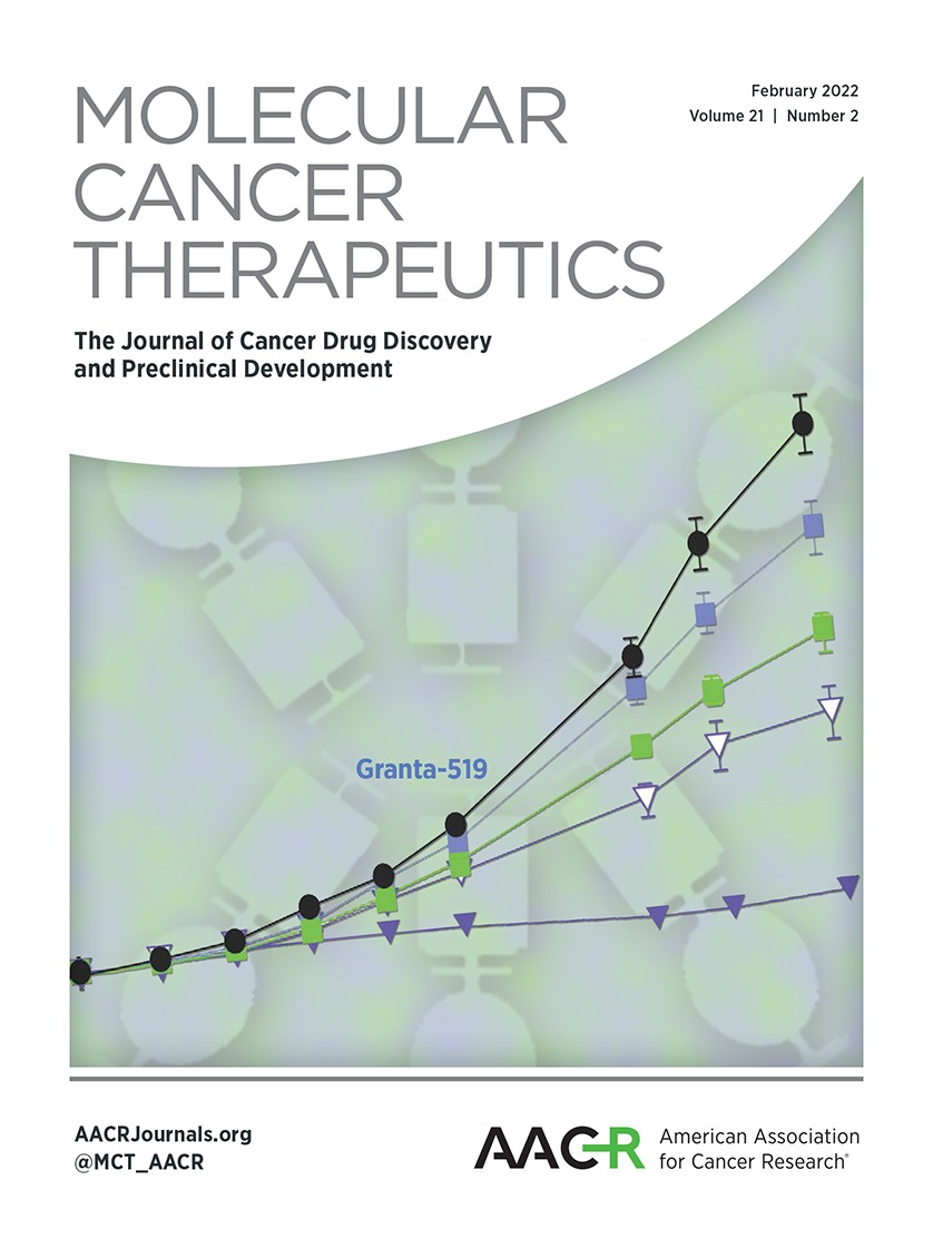
We next tested triple blockade of HER2, ER, and CDK4/6 in three HR+/HER2+ human breast cancer cell lines: BT474, MDA-MB-361, and UACC812. In all three cell lines, single agents tucatinib and fulvestrant had modest activity, whereas palbociclib had a greater tumoricidal activity at concentrations above 2.5 μmol/L, as would be expected from known therapeutic dosing in humans (Fig. 1A and B; Supplementary Fig. S2A). Responses to dual targeted combinations were similar between BT474 and UACC812, where tucatinib plus palbociclib and tucatinib plus fulvestrant reduced cancer cell viability better than single agents (Supplementary Fig. S2B and S2C). However, no dual combination was better than single agents in MDA-MB-361 (Supplementary Fig. S2D). The triple combination induced the most significant and consistent reduction in cancer cell viability across all three cell lines (Fig. 1A and B; Supplementary Fig. S2A).
 Figure 1.
 Activity of tucatinib, palbociclib, and fulvestrant in cancer cell lines. A–B, Triple combination improves suppression of tumor cell viability of (A) BT474 and (B) MDA-MB-361 cells. All cell line experiments were run in triplicate and repeated at least 3 times. Data are mean ± SEM. C–J, Signaling changes upon treatment with tucatinib (T), palbociclib (P), fulvestrant (F), or vehicle (V), treatment was conducted for 24 hours at the IC30 value: for MDA-MB-361, P = 1.9 μmol/L, F = 8.9 μmol/L, T = 0.05 μmol/L, for BT474, P = 1.5 μmol/L, F = 9.5 μmol/L, and T = 0.05 μmol/L. Average normalized expression via immunoblot of pRB S780 in (C) BT474 and (D) MDA-MB-361; pHER2 Y1221/1222 in (E) BT474 and (F) MDA-MB-361; ER in (G) BT474 and (H) MDA-MB-361; and pERK1/2 Y202/204 in (I) BT474 and (J) MDA-MB-361. Data are mean ±SD. Each western blot was done in triplicate and repeated with ≥2 sets of protein lysates. *, P ≤ 0.05 comparison between drug treatment and vehicle by the two-sided t test.
To further explore signaling changes upon targeted blockade, we selected BT474 and MDA-MB-361 cell lines (Fig. 1C–J; Supplementary Fig. S3A—S3N). As expected, in BT474 and MDA-MB-361, palbociclib suppressed phosphorylation of RB at serine 780 (pRB), a site that is phosphorylated only by the CDK4/6 complex (ref. 38; Fig. 1C and D; Supplementary Fig. S3A and S3H). Fulvestrant treatment resulted in some degree of pRB inhibition, consistent with published data that suppression of estrogen signaling may lead to inhibition of cell cycle and decreased pRB (39, 40). However, only treatment with palbociclib or palbociclib-containing drug combinations resulted in statistically significant decrease of pRB level compared with vehicle (Fig. 1C and D; Supplementary Fig. S3A and S3H). Tucatinib significantly decreased phosphorylation of HER2 in both cell lines (Fig. 1E and F; Supplementary Fig. S3C and S3J) and reduced pERK1/2 in BT474, but not in MDA-MD-361 (Fig. 1I and J; Supplementary Fig. S3E and S3L). When BT474 or MDA-MB-361 cells were treated with palbociclib, fulvestrant, or their combination, the level of pHER2 and pERK1/2 either did not change or increased; for example, fulvestrant increased pHER2 in BT474 (Fig. 1E), and both palbociclib and fulvestrant increased pERK1/2 in MDA-MB-361 (Fig. 1J). Dual combinations of tucatinib plus palbociclib or fulvestrant, or the triple combination, successfully suppressed pERK1/2 in both cell lines (Fig. 1I and J; Supplementary Fig. S3E and S3L). As expected, fulvestrant alone or in combinations decreased the level of total ER in both cell lines (Fig. 1G and H). Interestingly, tucatinib increased ER expression in BT474, and the same trend was noted in MDA-MB-361, although it did not reach statistical significance (Fig. 1G and H; Supplementary Fig. S3G and S3N). These results suggest that treatment with single-agent tucatinib may be insufficient to suppress downstream HER2 signaling; moreover, tucatinib may upregulate resistance pathways such as ER expression. In contrast, triple combination works along all three oncogenic pathways, resulting in reduction of pRB and ER expression, as well as complete suppression of HER2/pERK1/2 signaling, therefore, preventing potential escape mechanisms. In addition, our results suggest that the triple combination treatment may permit one drug to upregulate molecular targets for the other drugs in the combination.
Triple combination shows superior efficacy in suppression of tumor growth in vivoWe next tested our hypothesis that the combination of tucatinib, palbociclib, and fulvestrant would have efficacy in vivo. For in vivo experiments, we focused our analysis on the triple combination and dual combinations containing tucatinib (tucatinib plus fulvestrant or tucatinib plus palbociclib) and compared their activity with vehicle and single-agent tucatinib. We have elected not to test activity of single agents palbociclib, fulvestrant, or a combination of palbociclib and fulvestrant in animal experiments because published clinical studies have shown that HER2 inhibition is critical for treatment of patients with HER2+ breast cancer; the addition of HER2 inhibitors to combination treatment regimens improves OS (41, 42). Therefore, only tucatinib containing combinations are clinically relevant for patients with HR+/HER2+ disease.
MDA-MB-361 tumor growth was not reduced by tucatinib compared with vehicle (Fig. 2A). However, the combination of tucatinib with fulvestrant or palbociclib markedly reduced tumor growth, and the triple combination induced the most robust reduction in tumor growth. Average tumor volume at the EOT (VEOT) did not differ between tucatinib and vehicle groups, whereas VEOT in tucatinib plus palbociclib, tucatinib plus fulvestrant, and the triple combination groups were significantly smaller (Fig. 2B and C). VEOT on triple combination therapy was significantly reduced compared with both dual combinations (Fig. 2B and C). There was no difference in average TGR between vehicle and tucatinib; however, both dual combinations and the triple combination significantly reduced TGR (Fig. 2D). The TGR was the lowest in the triple combination, approximately 5.6 times lower than vehicle (Fig. 2D). IHC staining of Ki67 revealed that all palbociclib containing combinations had marked reduction of proliferation rate. However, the lowest percentage of proliferating cells was observed in the triple combination, where Ki67 was significantly lower compared with the best dual combination (Fig. 2E and F).
 Figure 2.
 Triple combination therapy reduces HR+/HER2+ tumor growth in vivo. MDA-MB-361 xenografts (N = 10–12 tumors per group) and BT474 xenografts (N = 9–12 tumors per group) were treated with vehicle (V), tucatinib (T) or combinations of tucatinib, fulvestrant (F), and palbociclib (P). Results shown for MDA-MB-361 xenografts: A, Tumor growth curves; B, Tumor volumes at end of treatment (EOT); C, Representative tumors at study completion (scale bar, 5 mm); D, Tumor growth rates (TGR; change in tumor volume over time) for each treatment group; (E) Ki67 analysis and (F) Images of representative Ki67 staining (scale bar, 25 μm). Results of BT474 xenografts are shown: G, Tumor growth curves; H, BT474 tumor volumes at EOT; I, Representative tumors from each treatment group at study completion (scale bar, 5 mm); J, TGRs; K, Ki67 analysis and (L) Images of representative Ki67 staining scale bar, 25 μm. All data are mean ± SEM. For B, D, E, H, J, and K overall differences are significant with P < 0.0001 by ANOVA; *, P < 0.05; **, P < 0.01; ***, P < 0.005; and ****, P < 0.001 by the two-sided t test
BT474 tumors yielded similar results where the triple combination induced the most robust reduction in tumor growth (Fig. 2G). Tucatinib alone or dual combination with fulvestrant or palbociclib also reduced tumor growth compared with vehicle treatments, although to a lesser extent than triple combination. Average VEOT in triple combination was significantly lower compared with tucatinib alone, tucatinib plus fulvestrant, or tucatinib plus palbociclib groups (Fig. 2H and I). The triple combination therapy was the only treatment where tumors were regressed in size as compared with the start of experiment. The triple combination induced a negative TGR, substantially lower than the TGR in the groups treated with single-agent tucatinib or tucatinib plus fulvestrant (Fig. 2J). Finally, Ki67 analysis revealed that all palbociclib containing combinations had marked reduction of proliferation rate, with the lowest Ki67 in the groups treated with palbociclib plus tucatinib or the triple combination (Fig. 2K and L). Full statistical analysis of animal experiments is summarized (Supplementary Fig. S4A). Mice did not experience significant weight loss on any treatment regimen, suggesting low toxicity of the triple combination in mammals (Supplementary Fig. S4B and S4C).
In vitro and in vivo experiments in drug-resistant cell linesWe next investigated potential cross-resistance mechanisms between two key components of the triple combination: tucatinib and palbociclib. We generated TR and palbociclib-resistant (PR) subclones of the BT474 and MDA-MB-361. We did not generate fulvestrant-resistant subclones, as all HR+/HER2+ cell lines exhibit intrinsic resistance to ER-targeting agents because of HER2 overexpression. We hypothesized that the mechanisms of resistance to tucatinib and palbociclib would be non-overlapping; therefore, cross-treatment of TR subclones with palbociclib and PR subclones with tucatinib would result in effective cancer cell killing.
In clonogenic assays, MDA-MB-361 TR and BT474 TR cells survived treatment with 0.67 μmol/L tucatinib; however, they had a significant reduction in clonogenic survival when treated with 3 μmol/L palbociclib (Fig. 3A and B). The addition of tucatinib to palbociclib did not improve reduction of clonogenic survival, reflecting profound resistance of TR subclones to tucatinib.
 Figure 3.
 HR+/HER2+ subclones resistant to tucatinib or palbociclib are sensitive to other agent in triple combination. Clonogenic survival of (A) BT474 and (B) MDA-MB-361 tucatinib-resistant (TR) subclones treated with vehicle (V), 0.67 μmol/L tucatinib (T), and/or 3 μmol/L palbociclib (P). Clonogenic survival of (C) BT474 and (D) MDA-MB-361 palbociclib-resistant (PR) subclones treated with vehicle, 10 μmol/L palbociclib, and 0.25 μmol/L (BT474) or 1.75 μmol/L (MDA-MB-361) tucatinib. E, Clonogenic survival of MDA-MB-361 PR subclones treated with vehicle, 5 μmol/L palbociclib, 1.5 μmol/L tucatinib, and/or 4 μmol/L fulvestrant (F). All clonogenic data are normalized to vehicle. All in vitro assays were replicated ≥3 times. Data are presented as mean ± SEM. F, Tumor growth curves of MDA-MB-361 TR tumors treated with vehicle or tucatinib. After tumor volumes reached 500 mm3, the tucatinib treatment group was switched to fulvestrant and palbociclib. N = 8–12 tumors per group. G, Representative tumors from vehicle and treatment groups at the end of the study. Dotted line represents length and width tumor volume measurements; scale bar, 1 cm. H, Representative Ki67 staining and (I) quantification; scale bar, 5 mm. *, P < 0.05; **, P <0.01; ***, P < 0.005; and****, P < 0.001 by ANOVA with Dunett multiple comparison test (A–E) or two-tailed t test (I).
Despite the resistance of BT474 PR subclones to palbociclib in normal cell culture growth conditions, they were partially sensitive to palbociclib in the clonogenic assay; yet, approximately 60% of cells survived palbociclib at 10 μmol/L. As predicted, BT474 PR cells were sensitive to tucatinib as evidenced by significantly reduced survival (Fig. 3C). Because BT474 PR cells retained partial sensitivity to palbociclib, the addition of tucatinib to palbociclib generated the most robust reduction in clonogenic survival. MDA-MB-361 PR cells were highly resistant to palbociclib, without any detectable suppression after treatment with 10 μmol/L of the drug. These cells were also relatively resistant to 1.75 μmol/L of single-agent tucatinib. Only treatment with both drugs resulted in significant reduction of clonogenic survival, yet almost 50% of cells survived treatment with tucatinib and palbociclib combination (Fig. 3D). To test whether the addition of the third agent could rescue the suppression of MDA-MB-361 PR cells that exhibited the phenotype of complete palbociclib and partial tucatinib resistance, we added 4 μmol/L of fulvestrant to treatment with 5 μmol/L palbociclib and 1.5 μmol/L tucatinib. The triple combination resulted in significantly lower survival than single agents (Fig. 3E).
Tucatinib did not significantly reduce growth of MDA-MB-361 TR mice xenograft tumors compared with vehicle, confirming that resistance to tucatinib is maintained in vivo (Fig. 3F). Once the tumors reached approximately 500 mm3, we adjusted the treatment regimen from tucatinib to a combination of palbociclib and fulvestrant, which substantially reduced growth of TR tumors compared with vehicle (Fig. 3F and G) and significantly reduced Ki67 positivity (Fig. 3H and I).
Although clonogenic survival assays assess the ability of single cancer cells to form a colony under stress, such as drug treatment (43), the CellTiter Glo assay measures cell viability in culture where cells are able to support each other via cell-to-cell contact. CellTiter Glo assessment of drug-resistant subclones on treatment with tucatinib, palbociclib, or fulvestrant alone, or in combination, confirmed that triple combination suppressed the viability of all drug-resistant subclones in MDA-MB-361 and BT474 cells (Supplementary Fig. Fig. S5A–SAD).
Triple combination–targeted therapy overcomes drug resistance mediated by cyclin EBecause the cyclin E–CDK2 complex is downstream of targets blocked by tucatinib, palbociclib, and fulvestrant, we were interested in evaluating cyclin E expression as a potential drug resistance mechanism (Fig. 4A). Cancer cells resistant to tucatinib or palbociclib increased expression of cyclin E in a stepwise fashion with increasing concentration of inhibitor in culture (Fig. 4B). In wild-type BT474 cells, cyclin E levels increased or remained the same on treatment with tucatinib, palbociclib, or fulvestrant, or their dual combinations; only the triple combination significantly reduced cyclin E (Fig. 4C; Supplementary Fig. S6A). In the wild-type MDA-MB-361 cells, multiple treatments reduced cyclin E expression, including single-agent palbociclib, palbociclib plus fulvestrant, tucatinib plus fulvestrant, and the triple combination (Fig. 4D; Supplementary Fig. S6B).
 Figure 4.
 Cyclin E expression as potential drug resistance mechanism. A, ER, HER2, and CDK4/6 signaling diagram and targeted agents. Diagram was created with BioRender.com. B, Cyclin E expression increases in BT474 palbociclib and TR subclones. Cells were treated with drug concentrations increasing in a step-wise fashion. Palbociclib 1.3 represents 1–1.49 range, 1.8 represents 1.5–1.99 range, 2.3 represents 2–2.49 range, and 2.8 represents 2.5–2.99 range. C, Cyclin E expression in wild-type (WT) BT474 and (D) MDA-MB-361 after treatment with vehicle (V), tucatinib (T), palbociclib (P), fulvestrant (F) or combination of drugs. All experiements were performed in triplicate with ≥2 sets of protein lysates; data are mean ±SEM; #, P ≤ 0.05 compared with WT by a two-tailed t test.
HR+/HER2+ breast cancers are enriched in young patientsAlthough the overall frequency of HR+/HER2+ disease is reported to be 10% of all breast cancer cases, we observe an enrichment of HR+/HER2+ disease in the University of Colorado Young Women's Breast Cancer Cohort among women diagnosed with breast cancer at the age of 45 or younger (Fig. 5A). Patients with HR+/HER2+ disease comprise 20% of this cohort, which was greater than the frequency of triple negative disease. Likewise, in The Cancer Genome Atlas, 20% of patients diagnosed with breast cancer at age 45 or younger had HR+/HER2+ disease (Fig. 5B), which was two times more common when compared with US SEER cohort comprised of all age patients (Fig. 5C).
 Figure 5.
 HR+/HER2+ breast tumors are more prevalent among young women. The percentage of patients with HR+/HER2+ breast cancer reaches 20% among women ≤45 years old in (A) the University of Colorado Young Women's Breast Cancer (YWBC) Cohort and (B) The Cancer Genome Atlas. C, The percentage of patients with HR+/HER2+ breast cancer is 10% in all age patients in the US SEER Database.
DiscussionHR+/HER2+ breast tumors represent a significant and under-recognized clinical problem. The simultaneous activity of ER and HER2 signaling provides multiple mechanisms for tumor survival and escape from therapy, enabling late recurrences and metastatic disease. We report that HR+/HER2+ tumors are enriched among young patients with breast cancer with a frequency similar to TNBC, and potentially contribute significantly to morbidity and mortality in this patient population, where breast cancer outcomes are disadvantageous compared with older patients (44). This is concordant with a previous reports that breast cancer in young women is enriched for luminal B tumor subtype (36, 45). Current clinical approach to treatment of HR+/HER+ breast cancer is not optimized. Ultimately, most patients with recurrent/metastatic disease experience resistance to the anti-HER2 monoclonal antibodies and ER-targeted therapies, and, as a result, are treated with chemotherapy-containing drug combinations. With the goal of developing a clinically effective targeted therapy approach, we tested a triple targeted blockade of HER2, ER, and CDK4/6 in HR+/HER2+ breast cancer cell lines and tumor xenograft models.
The key component of our combination therapy is tucatinib, a novel HER2 small-molecule inhibitor that, as we confirmed, is active in disease resistant to HER2-targeted antibodies (11–13). An additional benefit of tucatinib is its ability to prevent and treat brain metastases—a significant clinical advantage (13). With this newly available small-molecule inhibitor, we hypothesized that triple targeted therapy along the ER and HER2 pathways, along with CDK4/6 signaling inhibition, would overcome post-antibody therapy resistance and block the emergence of new treatment resistance as well. As expected, although dual combinations of tucatinib with palbociclib or fulvestrant were active in some of the cancer cell lines we studied, only the triple combination achieved consistent suppression of cell viability in all in vitro experiments. The triple combination achieved maximal suppression of tumor growth of BT474 and MDA-MB-361 xenografts, effectively eliminating the ability of tumor cells to proliferate in vivo and reducing Ki67 proliferation index to <1%. Notably, in both xenograft models, triple combination therapy resulted in the lowest VEOT that was significantly lower compared with dual combinations of tucatinib plus fulvestrant, and tucatinib plus palbociclib. Moreover, we demonstrated a TGR approaching zero in MDA-MB-361 xenografts, and a negative TGR in BT474 xenografts treated with the triple combination. It is well known that the ability of a treatment regimen to merely slow tumor growth is not sufficient for clinical benefit in humans; stable disease or partial responses are needed to demonstrate clinical activity in patients. Our in vivo experiments underscore high anti-tumor activity of triple combination and its potential to produce clinically meaningful responses. We acknowledge that this is the first in vivo study of the triple combination, and PDX experiments will be a logical next step in further studies. At the signaling level, our results suggest that treatment with the triple combination may permit one drug to upregulate molecular targets for the other drugs in the combination, which could explain high efficacy of the triple regimen. When all three drugs are applied together, cell cycle, ER, and HER2 pathways are consistently suppressed with no escape mechanism left along these pathways, which is not achieved by single agents or dual combinations.
Adaptive resistance of tumor cells to treatment is a cornerstone problem in medical oncology. Therapeutic approaches that initially inhibit tumor growth often fail due to adaptations of malignant subclones within heterogeneous tumors. Therefore, combination therapies with two or more agents designed to simultaneously target multiple pathways may be better suited to counteract tumor heterogeneity and adaptability. Palmer and Sorger postulated (46) that combination therapy approaches may be beneficial even without synergy between compounds, because each drug may be independently active against a certain population of drug-resistant cells. Using sophisticated mathematical modeling, Bozic and colleagues (47) demonstrated that dual combination approaches may result in better tumor control compared with single agents; however, triple combination therapy may be necessary for long-term prevention of drug resistance and durable responses in a clinical setting.
In concordance with the above, our experiments with drug-resistant subclones showed that triple combination is needed to acquire maximal benefit. Dual combination of palbociclib and tucatinib achieved some suppression of drug resistance, especially in the scenario of incomplete resistance to tucatinib or palbociclib. However, in our experiments, clonogenic survival and cancer cell viability were suppressed in only 3 out of 4 drug-resistant subclones, demonstrating that the mechanisms of cross resistance to tucatinib and palbociclib exist. It was only with the addition of the ER targeting agent (fulvestrant) that complete suppression was achieved. Because the subclones resistant to one agent may be effectively inhibited by the combination of the two remaining agents, the triple combination may be crucial to prevent the outgrowth of resistant subclones that could be either intrinsic or arising on therapy.
Finally, we report that expression of cyclin E oncogene increases in palbociclib and tucatinib resistant HR+/HER2+ cells, whereas the triple combination of tucatinib, fulvestrant, and palbociclib consistently reduces cyclin E expression in the two HR+/HER2+ cell lines that we tested. Cyclin E overexpression has been implicated in clinical resistance to HER2-targeted agents, CDK4/6 inhibitors, and anti-hormonal agents, and linked to adverse patient outcomes (18, 25, 26, 48). The ability of triple combination to overcome cyclin E-mediated drug resistance is potentially clinically valuable.
In summary, HR+/HER2+ breast cancers are particularly challenging to treat because of cross-talk between the HR and HER2 pathways, which promotes drug resistance and tumor progression. In addition, HR+/HER2+ breast cancers are inherently heterogeneous; approximately half of these tumors cluster as the HER2-enriched subtype, 24% cluster as luminal A, 20% cluster as luminal B, and 9% cluster as basal-like (49), which further contributes to treatment difficulty. Triple combination targeted therapy enables a more complete inhibition of tumorigenic signaling in a heterogeneous tumor cell population and can effectively counteract drug resistant subclones. This may solve an acute clinical problem of resistance to targeted agents, and result in more robust, long-term responses in patients. Our preclinical data, as well as available preliminary clinical data, support this promising regimen for further clinical trials (50).
Authors' DisclosuresE. Shagisultanova reports grants from Pfizer, Inc. and other support from Seagen during the conduct of the study. L.S. Crump reports grants from NIH during the conduct of the study. P. Kabos reports grants from Eli Lilly, Pfizer, AstraZeneca, Sanofi, Genentech, and Radius Health outside the submitted work. T.R. Lyons reports grants from NCI/NIH and ACS during the conduct of the study. V.F. Borges reports other support from SeaGen and grants from Pfizer during the conduct of the study; as well as other support from Olema, Cogent Therapetics, AstraZeneca, OncoSec, and Voyager Pharma outside the submitted work. No disclosures were reported by the other authors.
Authors' ContributionsE. Shagisultanova: Conceptualization, resources, formal analysis, supervision, funding acquisition, validation, investigation, visualization, methodology, writing–original draft, writing–review and editing. L.S. Crump: Formal analysis, visualization, writing–original draft, writing–review and editing. M. Borakove: Formal analysis, validation, investigation, methodology. J.K. Hall: Formal analysis, investigation, visualization. A.R. Rasti: Formal analysis, investigation, visualization. B.A. Harrison: Formal analysis, investigation, visualization. P. Kabos: Conceptualization, writing–review and editing. T.R. Lyons: Conceptualization, resources, formal analysis, funding acquisition, visualization, methodology, writing–review and editing. V.F. Borges: Conceptualization, resources, supervision, funding acquisition, methodology, project administration, writing–review and editing.
AcknowledgmentsThis work was funded by the NIH (KL2TR001080 and 1K08CA241071; to E. Sha
留言 (0)