
記住我

During the pandemic process of COVID-19, while the vaccination processes are ongoing, drug development studies that control the entry and replication of the SARS-CoV-2 into the cell will continue, and there will be discussions about whether it is good to suppress the uncontrolled immune response. This study aims to investigate the effects of whey proteins on SARS CoV-2 in methotrexate-induced lung tissue damage. Methotrexate (MTX) is an anti-metabolite of folic acid (vitamin B9) and, used in the treatment of cancer, autoimmune diseases, ectopic pregnancies, and psoriasis. It is also used for the treatment of pulmonary diseases such as pulmonary fibrosis. However, methotrexate can cause lung tissue damage which can lead to other complications such as pneumonitis and pulmonary fibrosis (COPD) (Koźmiński et al., 2020; Raimondi et al., 2019). It reduces thymidylate, purine synthesis, and cell proliferation by binding to reductase. It also inhibits folate-dependent enzymes resulting in adenosine accumulation and lymphocyte proliferation (Koźmiński et al., 2020). In patients under MTX treatment, the serum IL-1, IL-2, IL-6, IL-8, soluble IL-2 receptor (sIL-2R), and IL-1 receptor antagonist (IL-1RA) levels decreases (Mangoni et al., 2017; Sajjadi et al., 1996) and MCP-1 level does not change (Boiardi et al., 1999). While MTX treatment suppresses the immune system, therefore, the susceptibility to diseases increases in MTX using patients (Bedoui et al., 2019; Ibrahim et al., 2019). Caruso et al have shown that MTX prevents viral replication of the SARS COV-2 virus and the symptoms of COVID-19 (Caruso et al., 2021). SARS CoV-2 infection and lung cell destruction trigger a local immune response that recruits monocytes and macrophages to respond to infection, releasing cytokines, and setting up adaptive T and B cell immune responses, in most cases this process is capable to dissolve the infection. In some cases, however, a dysfunctional immune response occurs, which can lead to severe lung diseases. When the SARS-CoV-2 virus encounters a protective immune response in the host, there are marked reductions in COVID-19 symptoms (Tay et al., 2020; Wang et al., 2020). If the immune system cannot neutralize the effect of the virus, fatal complications can develop. Cytokine storm is one of these complications and is characterized as a dysfunctional immune response. High interleukin-6 (IL-6) level and high SARS-CoV-2 RNA level increase the lethal risk (Tay et al., 2020). Schalter et al found that angiotensin-converting enzyme-2 (ACE2) expression was decreased in the lungs, in the intestinal epithelium, and intestinal organoids of mice administered MTX (Schälter et al., 2021).
Furin (EC 3.4.21.85, proprotein convertase), is an enzyme in the mammalian subtilisin/kexin-like convertases. It activates several proproteins in all mammalian tissues (Nakayama, 1997). Lung alveoli epithelial cells exhibit furin at their plasma membrane. Furin and ACE2 are the receptors necessary for the SARS-CoV 2 entry into the cell (Johnson et al., 2020; Thomas, 2002). Furin cleaves the S1/S2 region of the spike protein of SARS-CoV-2, and the S1 unit binds tightly to the ACE2 receptor and enters the host cell. Although the S1/S2 region is cleaved by other proteases such as the transmembrane protease serine 2 (TMPRSS2) and cathepsin L, the pathogenicity of SARS CoV2 is high due to the high concentration of furin in tissues. The first step after the active site of SARS CoV2 existence is the binding of the virus to the host cell via the ACE2 receptor. Cheng et al. stated that furin inhibitors are potential candidates as an antiviral agent for COVID-19 treatment (Cheng et al., 2020).
Milk-derived caseins, whey proteins, tryptophan, lactadherin, mucin 1, lactoferrin, α-lactalbumin, and secretory immunoglobulin A (sIgA) are the potential candidates for the inhibition of SARS-CoV 2. (Roager & Licht, 2018). Among the milk-derived molecules, studies with whey proteins are at the forefront. Whey proteins play anti-inflammatory and immunostimulatory roles in metabolic syndromes. Whey proteins have been shown to improve lung function in patients with chronic obstructive pulmonary disease (Sugawara et al., 2012).
Wong et al demonstrated the increase of cell-mediated immune response with dietary whey proteins (Wong & Watson, 1995). Boukhettela et al showed the protective effect of whey proteins in the MTX-induced intestinal damage (Boukhettala et al., 2010). Recently Fan et al. (2020) have demonstrated the effects of breast milk whey proteins on viral infection and replication of SARS-CoV-2 in vitro. They also showed the inhibitor effect of commercial bovine formula milk on SARS-CoV-2 pseudovirus infection (Fan et al., 2020).
Therefore, this study aims to investigate the potential of whey protein concentrate to inhibit the activity of the lung furin enzyme and the binding of SARS-CoV 2 spike protein and ACE 2 in MTX-induced lung damage in rats.
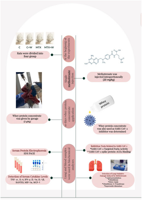
2 MATERIAL AND METHODS 2.1 Experimental designThe experimental protocols of this study were approved by the Marmara University School of Medicine Animal Care and Use Committee (Protocol code: 50-2021mar). Three-month-old male Sprague–Dawley rats weighing 200–300 g were used in the study. The rats were acclimatized at room temperature with a 12 hr light and dark cycle and fed with standard rat chow and had access to water ad libitum. Standard rat chow was purchased from MBD Feed Manufacturer, Kocaeli, Turkey. The standard rat chow contains 20% crude protein, 6% cellulose, 2.7% crude fat, 5.2% ash, 0.6% phosphorus, 0.18% sodium, and 0.115% vitamin mixture. Its metabolic energy was 2600 kcal/kg.
The animals were divided into four groups: six in the control group and eight in the other groups. MTX (David Bull Laboratories, Mulgrave-Victoria, Australia) was injected intraperitoneally (i.p). Whey protein beverage (Tazelen, Kaanlar Food Industry and Trade, Turkey) was administered to the rats by oral gavage. Following a single dose of methotrexate (in saline, 20 mg/kg; Mammadov et al., 2019), a whey protein beverage (2 g/kg) was administered for 10 days by gavage. On day 10, after decapitation of the rats, trunk blood was collected and, lung tissue was taken. Table 1 presents the experimental groups.
TABLE 1. Experimental groups Groups Saline (single dose, i.p) (0.1 ml/100 g) Methotrexate (single dose, i.p.) (20 mg/kg) Whey protein (10 days, oral) (2 g/kg) Saline (10 days, oral) (0.1 ml/100 g) C X – – X C + W X – X – MTX – X – X MTX + W – X X – Abbreviations: C, control (n = 6); C + W, control + whey protein (n = 8); i.p., intra peritoneal; MTX, methotrexate (n = 8); MTX + W, methotrexate + whey (n = 8); SD, standard deviation. 2.2 Preparation of whey protein concentrate (W)Whey protein beverage was lyophilized at −50°C under vacuum by using VIRTIS (SP Industries Inc., USA) freeze dryer. The protein concentration of the used whey protein beverage was 10 g %. Approximately 20 g whey protein powder was obtained after the lyophilization of a 50 ml whey protein beverage. The obtained whey protein powder was stored in the refrigerator until used. Whey protein powder was dissolved with tap water and was administered 2 g/kg to the relevant groups by oral gavage.
2.3 Preparation of serum samplesAfter taking the trunk blood samples, the blood was allowed to clot at room temperature for 15 min. After the clot was removed by centrifuging at 1,000×g for 10 min, the supernatant was taken to a new tube.
2.4 Analysis of inflammation in ratsSerum cytokine levels of the four groups were measured by the rat inflammation ELISA test (EA-1201, Signosis Inc., Sunnyvale, CA, USA). The test allows quantitative profiling and measuring eight inflammation cytokines (TNF- , IL-6, IFN-
 , IL-1α, IL-1β, MCP-1 [monocyte chemoattractant protein 1], CCL2 [chemokine ligand 2], RANTES [regulated on activation, normal T cell expressed and secreted], CCL5 [chemokine ligand 5], MIP-1α [macrophage inflammatory proteins], CCL3 [chemokine ligand 2]).
Electrophoretic examination of serum proteins was carried out by the principle of Laemmli SDS–polyacrylamide gel electrophoresis (SDS-PAGE) (Laemmli, 1970). SDS–PAGE was performed by using BIO-RAD mini protean precast II dual slab gel apparatus (BIO-RAD, USA). Mini PAGE gels (Any kD precast polyacrylamide gel, 8.6 × 6.7 cm [W × L], Catalog Number: 4569033, BIO-RAD, USA) were used for protein electrophoresis. 1:10 times saline diluted serum samples were used as a sample for SDS-PAGE.
2.6 Lung samplesLung tissues of all groups were homogenized with saline. Malondialdehyde (MDA) (Ledwozyw et al., 1986), glutathione (GSH) (Beutler, 1984), nitric oxide (NO) (Miranda et al., 2001), superoxide dismutase (SOD) (Mylroie et al., 1986), glutathione S transferase (GST) (Habig & Jakoby, 1981), and tissue factor (TF) (Ingram, 1976) activities were analyzed in 10% (wt/wt) lung homogenates. Lung homogenates were also used as a furin source for the furin inhibition test.
2.7 Determination of furin activity inhibitionThe lung furin activity targeting SARS-CoV-2 S1/S2 site cleavage was identified by the CoviDrop™ SARS-CoV-2 Targeted Proprotein Convertase Activity/Inhibition Assay Kit (Cat No: D-1007-48, Epigentek, USA). The in vivo inhibitor effect of whey protein on the lung furin activity was determined by using the lung tissues of each group as a furin source.
Two kinds of sample were used to determine the inhibition of lung furin activity performance of whey proteins.
Sample without inhibitor: The lung tissue of each group was accepted as furin source.
Sample with an inhibitor: The lung tissue of each group (furin source) and whey protein (inhibitor).
In this assay, polyhistidine and biotin tagged SARS-CoV-2 specific substrate was bound onto microplate wells. After the cleavage of the S1/S2 site, the S2 part of the substrate was removed by washing. The inhibitors that inhibit this cleavage prevented the signal reduction. The signal intensity was measured at a wavelength of 450 nm with an ELISA reader. The furin activity was inversely proportional to the signal intensity.
2.8 Determination of SARS-CoV-2 Spike-ACE2-binding activity inhibitionThe SARS-CoV-2 Spike-ACE2-Binding Activity Inhibition was determined by using the CoviDrop™ SARS-CoV-2 Spike-ACE2 Binding Activity/Inhibition Assay Kit (Epigentek, USA). In this inhibition test, the SARS-CoV-2 Spike-ACE2-binding activity inhibition percentage of whey protein concentrate, lung samples of all groups and serum samples of all groups were tested separately. In this test, the wells were coated with the SARS-CoV-2 spike protein. ACE2 of the sample or inhibitor was captured by the spike protein. The captured ACE2 amount was proportional to ACE2 binding activity. The absorbance was read at 450 nm in the ELISA reader. The intensity of the optical density was proportional to the ACE2 binding activity.
2.9 Statistical analysisGraphpad Prism 6.0 package program (GraphPad Software, San Diego, CA, USA) was used to evaluate the results. All data were presented as mean and standard deviation (SD). Analysis of variance (ANOVA) and followed by Tukey’s multiple comparison tests was used to compare the results of the groups. p < .05 was regarded as statistically significant.
3 RESULTS 3.1 Serum protein electrophoresisThe most significant change in serum protein electrophoresis results observed in the γ-fraction, which indicates the immunoglobulins. When the bands of the serum samples were evaluated visually, the serum γ-fraction of the MTX group was disappeared. Whey protein application enhanced visualization of the disappearing band (Figure 1).

Serum protein electrophoresis. C, control (n = 6); C + W, control + whey protein concentrate (n = 8); MTX, methotrexate (n = 8); MTX + W, methotrexate + whey protein concentrate (n = 8)
3.2 Cytokine levelsTNF-α, IL-6, and IL1-α levels were significantly decreased in the MTX group compared with the control group. It was determined that the administration of whey proteins to the MTX-administered group significantly increased these suppressed cytokines. IL-1β and MIP-1α levels were found to be significantly increased in the MTX group compared with the control group and significantly decreased with the administration of whey proteins compared with the MTX group (Figure 2). No significant difference was found between the IF-γ levels of all groups. MTX administration caused a significant increase in MCP-1 level compared with the control group, whey protein administration also significantly increased MCP-1 level both in C + W and MTX + W groups. While MTX administration did not significantly change the RANTES levels, the administration of whey proteins to the MTX group significantly decreased the RANTES levels compared with the other groups. While MTX administration significantly increased MIP-1α level, administration of whey proteins decreased MIP-1α level in both control and MTX groups.

Serum cytokine levels of all groups. (*): p < .05 compared with control group, (•): p < .05 compared with MTX group, (■): p < .05 compared with C + W group. C, control (n = 6); C + W, control + whey protein concentrate (n = 8); MTX, methotrexate (n = 8); MTX + W, methotrexate + whey protein concentrate (n = 8)
3.3 Lung resultsLPO level significantly increased in the MTX group compared with the control group. In the MTX group, the NO level significantly increased approximately three times the control group values. The GSH level, SOD and GST enzyme activities were also significantly decreased. Administration of whey proteins to the MTX group significantly decreased LPO and NO levels, significantly increased GSH level, SOD, and GST activity. The lung GST enzyme activity significantly increased to the control group activity with the administration of whey protein (Figure 3).

MDA, GSH, NO levels and SOD, CAT, and TF activities of lung tissue. (*): p < .05 compared with control group, (•): p < .05 compared with MTX group, (■): p < .05 compared with C + W group. C, control (n = 6); CAT, catalase; C + W, control + whey protein concentrate (n = 8); GSH, glutathione; MDA, malondialdehyde; MTX, methotrexate (n = 8); MTX + W, methotrexate + whey protein concentrate (n = 8); NO, nitric oxide; SOD, superoxide dismutase; TF, tissue factor activity
3.4 SARS CoV-2-targeted lung furin activity inhibitionTable 2 presents the inhibition percentages of lung furin activity targeting SARS-CoV-2 S1/S2 site cleavage by whey protein in each group. When the whey proteins were used as an inhibitor, the percentage of lung furin activity inhibition of the C + W group was significantly higher than the C group. Furin activity inhibition of the whey protein was significantly higher in the lungs of MTX-treated rats compared with the control group. When the whey protein was used as an inhibitor, the percentage of lung furin activity inhibition of the MTX + W group was found to be significantly lower than the furin activity inhibition of the C + W and MTX groups and significantly higher than the C group (Table 2).
TABLE 2. Lung furin activity inhibition of all groups with whey protein concentrate Lung C C + W MTX MTX + W Mean SD Mean SD Mean SD Mean SD Furin activity inhibition (%) 33.4 3.2 83.7* 4.6 70.1*,■ 5.7 52.4*,•,■ 3.4 Note (*): p < .05 compared with control group, (•): p < .05 compared with MTX group, (■): p < .05 compared with C + W group. Abbreviations: C, control (n = 6); C + W, control + whey protein concentrate (n = 8); MTX, methotrexate (n = 8); MTX + W, methotrexate + whey protein concentrate (n = 8); SD, standard deviation. 3.5 SARS-CoV-2 spike-ACE2-binding inhibitionSARS-CoV-2 Spike-ACE2-binding activity inhibition percentage of whey protein alone was found at 57%. Spike-ACE2-binding activity in serum and lung samples of the control group and serum samples of the MTX group was under the detection limit. The SARS CoV-2 Spike-ACE2-binding inhibition percentage of lung and serum samples of the control group was under the detection limit. Administration of whey proteins to the control group significantly increased the SARS CoV-2 Spike-ACE2 binding inhibition percentage of lung and serum samples. The SARS CoV-2 Spike-ACE2 binding inhibition percentage of serum samples of the MTX group was under the detection limit. Administration of whey proteins to the MTX group significantly increased the SARS CoV-2 Spike-ACE2-binding inhibition percentage of serum sample and significantly decreased the SARS CoV-2 Spike-ACE2-binding inhibition percentage of the lung samples. Control and methotrexate group. The lowest SARS CoV-2 spike-ACE2-binding inhibition was detected in the lung of the MTX + W group, the highest inhibition was detected in the serum sample of the C + W group (Table 3).
TABLE 3. SARS-CoV-2 Spike-ACE2-binding activity inhibition of lung and serum samples of all groups SARS-CoV-2 spike-ACE2 binding activity inhibition C C + W MTX MTX + W Mean SD Mean SD Mean SD Mean SD Lung (%) Under detection limit 51.9* 5.4 40.8* 4.3 23.6*,•,■ 2.4 Serum (%) Under detection limit 58.5* 3.2 Under detection limit 49.6*,•,■ 3.1 Note (*): p < .05 compared with control group, (•), p < .05 compared with MTX group, (■): p < .05 compared with C + W group. Abbreviations: C, control (n = 6); C + W, control + whey protein concentrate (n = 8); MTX, methotrexate (n = 8); MTX + W, methotrexate + whey protein concentrate (n = 8); SD, standard deviation. 4 DISCUSSIONIn this study, the effects of whey proteins on MTX-induced lung damage were investigated. Since the lung is the organ affected by the COVID-19, the inhibitory effects of whey proteins on the lung furin activity in MTX-induced lung damage and also on the SARS CoV-2 spike protein and ACE2 binding were also investigated. The detection of the effects of whey proteins on MTX-induced lung damage was significant for associating these effects with the parameters related to SARS CoV-2.
Although methotrexate is generally considered safe and easy to use, it has some side effects. Lung toxicity is a well-described side effect of methotrexate and can take a variety of forms. Pulmonary infiltrates are the most common form of methotrexate-induced lung toxicity, and these infiltrates are similar to hypersensitivity lung disease (Lateef et al., 2005). Kishta et al stated that the use of substances of nutritional origin with anti-inflammatory and antioxidant properties, such as whey proteins, makes it possible to limit bacterial infection of the respiratory tract, in particular, under conditions of permanent oxidative stress (Kishta et al., 2013). Garg et al showed that whey protein concentrate supplementation can restore the redox state in erythrocytes and thus maintain membrane transporter activity during ageing in rats (Garg et al., 2018). In the present study, the administration of whey proteins improved the MTX-induced lung damages in terms of LPO and GSH levels, SOD, GST, and TF activities. The lung oxidative balance, which is disrupted by MTX administration, may be further disrupted by COVID-19-based oxidative stress. Although there are no studies in the literature linking lung tissue damage caused by COVID-19 and MTX, the new preclinical evaluation of MTX for the treatment of COVID-19 has not been successful (Stegmann et al., 2021). SARS CoV-2 uses the human angiotensin I converting enzyme 2 (ACE2) for entry into the cells with its spike protein. ACE2 is expressed in the epithelial cells of the lung and other organs. ACE2 was recently identified as the receptor for SARS CoV-2, the infectious agent responsible for COVID-19, which provides a critical link between immunity, inflammation, ACE2, and cardiovascular disease (Gheblawi et al., 2020; Kishk et al., 2021; Zimmerman et al., 2004).
Numerous studies have been carried out to investigate the effects of various proteins on the SARS CoV-2 infection. Whey protein supplementation represents a practical, feasible, and cost-effective approach to alleviating malnutrition, high cholesterol, tumor cachexia syndrome, and hypertension (Bumrungpert et al., 2018; Fekete et al., 2016; Stobaugh, 2018). Therefore, in this study, the potential of whey proteins to protect the lung against SARS CoV-2 in MTX-induced lung injury was determined. The usefulness of whey protein is evidenced by a higher leucine content and the potential to modulate IGF-1 concentrations, which are necessary factors for musculoskeletal hypertrophy (Teixeira et al., 2019). Fan et al. (2020) observed that whey protein from unique species such as cow and goat has anti-coronavirus properties, and industrial bovine milk also confirmed comparable activity. They additionally showed anti-microbial contents of breast milk, such as lactoferrin and IgA antibody, that have limited anti-coronavirus activity, suggesting that breast milk could play the anti-coronavirus role. Boukhettela et al have revealed that a specific diet containing whey proteins has a protective effect in rats given repeated courses of MTX by improving nutritional outcomes and reducing intestinal damage. (Boukhettala et al., 2010). Çakır et al presented the inhibitory roles of beta-lactoglobulin and its peptides, one of the whey proteins, on SARS CoV-2 by in silico analysis (Çakır et al., 2021). In the present study, when whey protein was used as an inhibitory agent and the MTX-treated lung was used as a furin source, the lung furin inhibition of the MTX group was higher than the C and MTX + W groups. The lung furin inhibition of the MTX + W group was also higher than the C group. Stegman et al revealed that SARS CoV-2 replication was inhibited by MTX in therapeutic concentrations of 1 μM, which led to a more than a 1000-fold decrease in virus progeny in Vero C1008 cells and Calu3 cells. They also stated that the inhibitory effect of MTX was primarily based on reduced purine synthesis (Stegmann et al., 2021). When whey protein was used as an inhibitor of furin activity and lung of group C + W was used as a source of furin, inhibition of furin was higher in the lung of group C + W compared with the C group. This finding showed that whey protein inhibited lung furin activity of the C + W group more effectively than C group. Therefore, the cleavage of SARS CoV-2 into its subunits in the lungs of the C + W group is further inhibited so that SARS CoV-2 did not bind to its receptors in the lung of the C + W group compared with the C group. In this study, MTX caused oxidative damage in the lung tissue and activated the inhibitor mechanisms against SARS CoV-2. While the administration of whey proteins to the MTX group improved oxidative damage in the lung tissue, it impaired the inhibitor mechanisms against SARS CoV-2, but this effect was still higher than the control group that did not receive any whey protein. Although a detailed clinical study is needed, this finding can be associated with the fact that healthy people using whey protein before the onset of COVID-19 may obtain more inhibition than when they start using whey protein in the case of COVID-19. The second inhibition test related to SARS CoV-2 in this study was the inhibition of SARS CoV-2 spike protein and ACE 2 binding. According to the results of this study, whey protein alone inhibited the SARS-CoV-2 Spike-ACE2 binding activity by 57%. No SARS-CoV-2 Spike-ACE2 binding inhibition was detected when the lung and blood samples of the control group were used as an inhibitor. Administration of whey protein to the control group increased this inhibition approximately 50%–60% when lung and blood samples of this group were used as an inhibitor. While the spike protein and ACE 2 binding was inhibited by approximately 40% when lung tissues of the MTX group was used as an inhibitor, no such inhibition was detected when the blood samples of this group was used as an inhibitor. Administration of whey protein to the MTX group increased the inhibition of spike protein and ACE2 binding approximately 50% when blood sample of this group was used as an inhibitor. This inhibition can be associated with the whey protein-based peptides found in the blood of the MTX + W group. These results indicate that whey protein treatment may suppress the SARS-CoV-2 infection by inhibiting the lung furin activity and spike protein-ACE2 binding.
In this study, MTX administration also decreased serum TNF-α, IL-6 and IL-1α levels, increased serum IL-1β, MCP1 and MIP-1α levels and did not change serum IF-γ and RANTES levels compared with the control group. These findings are consistent with studies in the literature (Kim et al., 2009; Olsen et al., 2014). Giving whey protein to MTX-treated animals reversed all these changes and also caused a decrease in RANTES level but not in IF-γ level. Additionally, the decreased serum γ-globulin fraction detected by SDS–PAGE in the MTX-treated group also reflects changes in the immunoglobulin profile. The administration of whey proteins restored the changed γ-globulin fraction profile similar to the control group.
In conclusion, MTX-induced oxidative lung injury was ameliorated by whey proteins. By giving whey protein to the MTX-treated rats, lung tissue improved and, the protection against SARS CoV continued, albeit at a reduced rate. In healthy rats, 10 days of whey protein administration (C + W group) showed higher inhibition on the furin activity and the spike-ACE2 binding when compared with the control group that was not administered whey protein. Whey proteins belong to the protein class that deserves clinical investigation in terms of both MTX-induced lung damage and its potential to inhibit SARS-CoV2-related interactions.
CONFLICT OF INTERESTSThe author declares that there is no conflict of interest that could be perceived as prejudicing the impartiality of the research reported.
AUTHOR CONTRIBUTIONSElif Tufan: Investigation; Methodology. Güzin Göksun Sivas: Investigation; Methodology. Begüm Gürel-Gökmen: Investigation; Methodology. Sümeyye Yılmaz-Karaoğlu: Investigation; Methodology. Dursun Ercan: Investigation; Methodology. Dilek Özbeyli: Investigation; Methodology. Göksel Şener: Conceptualization; Investigation; Methodology; Supervision. Tuğba Tunali-Akbay: Conceptualization; Methodology; Project administration; Supervision; Validation; Writing – original draft; Writing – review & editing.
ETHICAL APPROVALThe experimental protocols of this study were approved by the Marmara University School of Medicine Animal Care and Use Committee (Protocol code: 50-2021mar).
留言 (0)