
記住我

Multiple sclerosis (MS) is a disabling autoimmune disease of the central nervous system (CNS) characterized by demyelination and neurodegeneration.1 It affects approximately 2.5 million people worldwide and poses a growing burden to society.2, 3 Relapsing-remitting MS (RRMS) is the most common initial course featuring alternate relapse and remission, and disability is aggravated gradually with illness development.4 After approximately 20 years, around 90% of RRMS patients will develop secondary progressive MS (SPMS) characterized by progressive neurodegeneration without any definite remission periods.5, 6 In addition to SPMS, progressive MS (PMS) also includes primary progressive MS featuring inapparent clinical relapses from the onset. At present, both immune and nonimmune mechanisms are believed to be involved in MS pathogenesis.
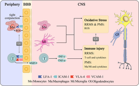
The “outside-in” hypothesis proposes that the inflammatory demyelinating process begins in the subarachnoid space and cortex and extends into the white matter.7, 8 In this model, the invasion of peripheral immune cells disrupts the blood-brain barrier (BBB) integrity and contributes to the prolonged presence of inflammatory activity. In RRMS, the interaction of monocytes and brain endothelial cells (ECs) produces massive reactive oxygen species (ROS), leading to the loss of tight junctions and migration of monocytes.9 For T cells, the mutual recognition of lymphocyte function-associated antigen-1 (LFA-1), intercellular cell adhesion molecule-1 (ICAM-1), very late antigen-4 (VLA-4), and vascular cell adhesion molecule-1 (VCAM-1) permits them to cross the BBB. The release of matrix metalloprotein-9 (MMP-9) by T cells is also essential for the migration process. Notably, infiltrated T cells can recruit macrophages, microglia, and astrocytes by secreting mediators, including tumor necrosis factor-α (TNF-α), interferon-γ (IFN-γ), and interleukins-17 (IL-17).10, 11 These abnormally activated immune cells target neurons and the myelin sheath and drive MS relapse and progression. Therefore, several disease-modifying therapies (DMTs) can decrease relapse rates by immunomodulation. However, the potential risks of serious adverse events (AEs) and COVID-19 infection limit its clinical use to some extent.12, 13 In PMS, inflammation is compartmentalized and mainly driven by the activities of innate microglia, astrocytes, and B cells.11 Unfortunately, the efficacy of DMTs for PMS tends to be disappointing, motivating the search for a new treatment option.14
Oxidative stress is another crucial driver of MS once the autoimmune system has caused damage to the CNS.10 It occurs when an imbalance exists between excessive production of free radicals and insufficient biological ability to remove them.15 The CNS is quite sensitive and vulnerable to oxidative stress because of its high oxygen consumption and lipid abundance. Oxidizing substances, such as ROS and nitrogen species, are usually produced by activated macrophage and microglial structures, causing damage to lipids, proteins, and DNA. Consequently, the CNS is variously disrupted through processes such as increased BBB permeability, myelin phagocytosis, and neurodegeneration.16, 17 In the plasma of MS patients, the levels of antioxidants and total antioxidant capacity are decreased.18, 19 Autopsy studies have also widely detected the damage induced by oxidative stress in cerebrospinal fluid and CNS tissues.20, 21 Therefore, oxidative stress may be another hopeful therapeutic target of MS. At present, many antioxidant compounds have improved serological indicators in MS patients.22 Vitamin D decreased the relapse rates as an antioxidant in RRMS patients.23 However, the findings of the efficacy of antioxidants tend to be conflicting and confusing, strongly suggesting that the effect of using a single antioxidant is limited. Considering the above, an ideally effective medicine must possess the ability to prevent multiple pathogenic factors and outstanding BBB permeability.
Lipoic acid (LA), also known as thioctic acid, has become a hopeful complementary therapy in MS to target both inflammation and oxidative stress. LA is a double-sulfhydryl natural antioxidant with two enantiomers according to optical rotation: R-LA and S-LA. Overall, R-LA exists widely in plants and animals, whereas S-LA is artificially synthesized to compose the racemic mixture (1:1 R/S-LA).24 In the human body, R-LA is synthesized de novo by cysteine and fatty acids in small amounts; thus, it primarily depends on exogenous supplements such as organ meat, broccoli, and fruits.25 For individuals, the racemic form can be absorbed rapidly after oral administration and participate in various biological metabolic pathways. First, it contributes to the synthesis of vitamin C and vitamin E.26 Second, it is reduced to dihydro-LA (DHLA), and DHLA is involved in the biosynthesis of intracellular glutathione (GSH) and coenzyme Q10.27, 28 Third, R-LA plays a crucial role in mitochondrial energy production as a cofactor for some enzymatic complexes in the Krebs cycle.29 When other metabolic pathways are saturated, redundant LA (nearly 10%) will be excreted through the kidneys.30 Over the past two decades, whether LA improves the quality of life of patients with MS has been intensively studied.31 In mouse models of experimental autoimmune encephalomyelitis (EAE), LA increased the population of mature oligodendrocytes and alleviated neurological symptoms, suggesting that LA might protect and promote neuronal regeneration.32, 33 However, the results of alleviated neurological symptoms were inconsistent for different administration pathways, timing, and dosage, making the evidence somewhat fragile. In patients with MS, LA reduced the Expanded Disability Status Scale (EDSS), although the between-group difference was not statistically significant.34, 35 The confusing result regarding whether LA could improve patient outcomes probably resulted from the short trial duration. Additionally, the annualized percent brain volume change was less after 2 years of supplementation in the LA group, indicating that LA might prevent neuronal death and reduction.36 More importantly, few AEs were reported when using LA as an oral preparation for 2 years. In summary, LA shows strong antioxidative and anti-inflammatory effects in MS, which makes it a potential candidate for complementary and long-term therapy.
To date, no study has systematically summarized the current findings of LA in MS, and some results appear to be controversial. A good review of both achievements and limitations will contribute to determining reliable evidence and research trends for future studies. In this review, we aimed to provide comprehensive insight into the role of LA in MS, including the aspects of pharmacokinetics, efficacy, safety, and mechanism, in both in vitro and in vivo experiments. We hope that our work will contribute to the development of new drugs and combination therapy for patients with MS.
2 METHODS 2.1 Search strategyAccording to the guidelines of the 2009 Preferred Reporting Items for Systematic Reviews and Meta-Analysis (PRISMA) statement,37 English-language studies published from inception up to July 1, 2021, were collected by searching five databases: PubMed (Medline), EMBASE, Web of Science, Scopus, and Cochrane Library. Identified search terms included (“multiple sclerosis” OR “MS”) AND (“lipoic acid” OR “Thioctacid” OR “LA”). Additional records were identified manually through other sources, such as any related review papers and reference lists of all included studies to avoid missing relevant studies in the initial search. The whole search process was conducted by two authors independently (H.S.X. & X.F.Y.).
2.2 Study selectionAfter removing duplicates, all the studies were screened for eligibility by two independent authors (H.S.X. & X.F.Y.). The inclusion criteria in this systematic review included the following: (1) randomized intervention study in patients meeting the McDonald criteria for MS38, 39; (2) preclinical experiments based on the mouse and cell models of MS; and (3) publications in peer-reviewed journals. The exclusion criteria included the following: (1) combined other antioxidants; (2) irrelevant endpoints such as biochemical metabolism and visual changes; and (3) nonoriginal studies. All the included studies were cross-checked, and in-depth discussions were required to resolve disagreements and make the ultimate decision with the senior author (Z.Y.J.).
2.3 Data extractionFor the included studies, we collected the following information into a spreadsheet in Excel: (1) subject characteristics including age, sex, EDSS score, and MS duration; (2) MS-related model establishment in the preclinical experiments; (3) LA dosage (4) endpoints including efficacy, safety, pharmacokinetics, and mechanism; and (5) first author's name, publication date, study design, and follow-up duration. For detailed data not shown in the full text, the e-mails were sent to the corresponding authors for help.
3 DISCUSSIONWe obtained 516 potential records in the initial systematic search. After the removal of duplicates, 143 studies were screened based on the title and abstract, leading to 59 full-text studies screened for eligibility. In this process, 27 articles were excluded because of irrelevant endpoints, nonoriginal studies, and combined antioxidant supplements. Finally, 32 intervention studies were included in this systematic review to investigate the effects of LA on efficacy, safety, pharmacokinetics, and mechanism. An overview of the study selection is presented in Figure 1.

Preferred Reporting Items for Systematic Reviews flowchart
3.1 LA pharmacokinetics and transportation to the brainA rat experiment found that the duodenum was the best portion of the intestine for LA absorption and that R-LA showed a higher absorption percentage than S-LA.40 Notably, two vital pathways are involved in the process of LA crossing the intestinal barrier: Na+/multivitamin (SMVT) and monocarboxylic acid (MCT) transporters.41, 42 Under equilibrium conditions, human SMVT can simultaneously bind and transport two LA molecules into the mesenteric vein, and human MCT transports LA in an energy- and low-pH-dependent manner. In the patients with RRMS/SPMS and healthy volunteers, the pharmacokinetic parameters showed no significant difference, suggesting that the MS status did not influence LA metabolism.43 In 54 patients with SPMS, pharmacokinetics showed no significant difference between the baseline and 1 year later, suggesting that the oral administration of LA was stable for long-term use.44 In patients with MS, three studies found that the time to reach the peak concentration of R-LA was much shorter than that of the racemic form, indicating the quicker absorption of the R-configuration.33, 45 Additionally, R-LA showed a much larger area under the curve than the racemic form under the same dosage, indicating the better utilization of the R-configuration. In summary, R-LA showed quicker and better utilization than the racemic form, emphasizing the necessity of unified formulations and encapsulations if considering it as a supplementary therapy.
Notably, human SMVT and MCT are also expressed in brain microvessels and contribute to the transportation of LA across the BBB.46, 47 In an in vitro experiment, LA showed the ability to cross the BBB and exert beneficial effects on the viability of astrocytes.48 Besides, a rat experiment found that14C-labeled LA reached peak levels in the cortex, spinal cord, and sciatic nerve after one-half hour of oral administration, indicating that LA was taken up by both the CNS and peripheral nerves.49 LA was also measured in the rat brain cortex, cerebellum, striatum, and hippocampus after intravenous and intraperitoneal administration.50, 51 Notably, a recent rat experiment found that the LA did not cross the BBB as easily as supposed after the correction for blood volume, which emphasized that the permeability of the BBB might be greatly influenced by cerebral blood flow.52
3.2 Role of LA in cell experimentsHuman peripheral blood mononuclear cells (PBMCs) are isolated from peripheral blood and feature round nuclei. They mainly comprise lymphocytes, monocytes, and NK cells.53 Most PBMCs are naïve without immune effects. Importantly, the largest fraction, T cells, will develop into diverse subsets of Th1, Th2, Th17, or regulatory T cells (Treg cells) after activation by different cytokines.54, 55 Monocytes in PBMCs can also be activated by proinflammatory factors to simulate the immune status of MS. These make human PBMCs a suitable model of MS and provide an opportunity to mirror the autoimmune response in the CNS. Additionally, murine cell models of MS are established directly by isolating and culturing brain cells, including primary microglial cells stimulated with lipopolysaccharide/IFN-γ and primary cortical neurons treated with H2O2. We included nine studies based on human PBMCs or murine cells, which were treated with 10–100 μg/ml LA (Table 1). No study reported the specific form of LA, and only one study indicated the usage of both LA and DHLA.
TABLE 1. Effects of LA on MS in the preclinical studies Study Subjects LA dosage Antioxidation Immunomodulation Neuroprotection Duration Sanadgol et al32 36 mice 20–40 mg/kg LA, ip ROS (–) NA OLG (+) Bax/Bcl−2, caspase−3 (–) 5 weeks Yadav et al33 49 mice 5–100 mg/kg R/S LA, ih NA NA 10-Day CDS (–) 7 weeks Marracci et al56 Human T-cell 50–100 μg/ml LA 25–100 μg/ml DHLA NA T-cell migration (–) VLA−4, MMP−9 (–) NA NA Salinthone et al. 201057 Human PBMC 50–100 μg/ml LA NA T-cell proliferation (–) IL−6, IL−17 (–) cAMP (+) NA Lee et al58 Human monocyte 250 mmol/l LA NF-κβ (–) ICAM−1 (–) NA NA George et al59 Human PBMC 100 μg/ml LA NA Monocyte migration (–) B-cell migration (–) NA NA Salinthone et al60 Human PBMC 10–100 μg/ml LA NA NK-cell cytotoxicity (–) INF-γ (–) NA NA Fiedler et al61 Human PBMC 25–100 μg/ml LA NA Phagocytosis (–) IL−1β (–), cAMP (+) TNF-α, IL−6 (=) NA NA Schillace et al62 Human PBMC 100 μg/ml LA NA NA cAMP (+) NA Chaudhary et al63 Murine microglia 25–100 μg/ml LA NA Phagocytosis (–) NA NA Barsukova et al64 Murine neurons 100 μg/ml LA ROS (–) NA Axonal integrity, cAMP (+) NA Marracci et al69 87 mice 10–50 mg/kg R/S LA, sc NA T-cell, MMP−9 (–) 10-Day CDS (–) 7 weeks Morini e70 45 mice 5 mg/kg LA, orally 50 mg/kg LA, ip NA Immune infiltration (–) INF-γ, IL−4, MMP−9 (–) Disease scores (–) 6 weeks Schreibelt et al. 2006 71 14 rats 10–100 mg/kg R/S LA, sc NA Monocytes (–) Clinical signs (–) BBB permeability (–) 3 weeks Wang et al72 20 mice 50 mg/kg LA, injection PPAR-γ (+) Immune infiltration (–) Tregs (+) Clinical score (–) 3 weeks Li et al73 Mice 100 mg/kg LA, ip SOD (+) Malondialdehyde (–) Immune infiltration (–) TNF-α (–), Tregs (+) Clinical signs, demyelination (–) Axons (+) 26 weeks Dietrich et al74 Mice 100 mg/kg R/S LA, orally Glutathione (+) Immune infiltration (–) Disability score (–) RGC (+) 17 weeks Khan et al75 24 mice 3–10 mg/kg LA, sc NA Immune infiltration (–) Neuropathic pain (–) 5 weeks Chaudhary et al81 12 mice 100 mg/kg LA, sc NA Immune infiltration (–) ICAM−1, VCAM−1 (–) NA 3 weeks Chaudhary et al82 40 mice 100 mg/kg LA, sc NA Immune infiltration (–) NA 3 weeks Note “-” indicates reduced expression or inhibited activity compared with non-LA group, and “+” indicates increased expression or enhanced activity. Abbreviations: BBB, Blood-brain barrier; cAMP, Cyclic adenosine monophosphate; CDS, Cumulative Disease Score; COX-2, Cyclooxygenase-2; ICAM-1, Intercellular cell adhesion molecule-1; Ih, Subcutaneous injection; INF-γ, Interferon-γ; Ip, Intraperitoneal injection; MMP-9, Matrix metalloprotein-9; NA, Not available; OLG, Oligodendrocytes; PBMC, Peripheral blood mononuclear cells; PGE2, Prostaglandin E2; PPAR-γ, Peroxisome-proplator-actified receptor-γ; RGC, Retinal ganglion cells; ROS, Reactive oxygen species; Sc, Intramuscular injection; SOD, Superoxide dismutase; TNF-α, Tumor necrosis factor-α; VCAM-1, Vascular cell adhesion molecule-1; VLA-4, Very late antigen-4.Overall, LA inhibited the expression of various inflammatory mediators and the activities of immune cells in human PBMCs. In human T cells, LA inhibited cellular transmigration across a fibronectin barrier in a dose-dependent manner, likely because LA could downregulate the surface expression of VLA-4 and decrease the MMP-9 content in culture supernatants.56 Additionally, the oral administration of LA inhibited T-cell proliferation and activation enriched from the PBMCs of MS patients, which might be related to elevated intracellular cyclic adenosine monophosphate (cAMP).57 Further investigation demonstrated a lower content of IL-6 and IL-17 in culture supernatants than that in the non-LA group. In human monocytes, LA inhibited cellular migration in a dose-dependent manner.58, 59 Importantly, this effect might be related to the reduced activity of nuclear transcription factor-kappa B (NF-KB), leading to the decreased expression of TNF-α, MMP-9, and ICAM-1.31 LA also lowered the percentage of phagocytic cells in a dose-dependent manner in monocytes from both healthy controls and patients with RRMS. In human PBMCs, LA decreased the expression of various proinflammatory cytokines, including IL-1β, IL-6, IL-17, and IFN-γ.60, 61 However, some studies reported no difference in expression of TNF-α and IL-1β between the LA and non-LA groups, which may be explained by the modeling approach of lipopolysaccharides and the relatively small sample size. Notably, three studies revealed that the above anti-inflammatory and neuroprotective effects might be closely associated with elevated intracellular cAMP expression.62 Notably, the above outcomes based on PBMCs should be interpreted carefully because they lack in vivo environmental stimuli.
In murine cell models of MS, LA protected neurons and disturbed the activities of immune cells. In murine IFN-γ-activated microglia, LA disorganized the actin protein and disturbed the formation of membrane blebs, likely leading to alterations in cellular mobility and phagocytosis.63 In murine H2O2-treated cortical neurons, oxidative stress led to a marked increase in axoplasmic Ca2+ and the formation of the axonal spheroid, where axonal severing occurred. Interestingly, pretreatment with LA completely prevented spheroid formation and maintained axonal integrity by increasing the levels of cAMP.64 Presently, the main limitation of cell experiments resides in the nonunified methods of establishing the MS model. CNS cells of EAE mice are likely the most appropriate and convincing model for the double hit of autoimmunity and oxidative stress.
3.3 Role of LA in animal experimentsEAE is a reliable murine model that can well simulate the occurrence and development of MS.65 In the 11 included studies, EAE induction was accomplished using three methods (Table 1). Five studies indicated that the mice were immunized according to the standard protocol with complete Freund's adjuvant (CFA) containing oligodendrocyte glycoprotein fragment 35–55 or guinea pig myelin basic protein. Four studies indicated that the mice were immunized using CFA containing proteolipid protein (PLP) 139–151 peptide. PLP is a hydrophobic integral membrane protein accounting for half of the protein content of CNS myelin and was recently proven to correlate with the severity of disease in MS patients.66 In addition to the autoimmune component, the oral administration of cuprizone can cause whole-brain demyelination and gliosis and was used to establish the murine model in one study. Compared with the two previous methods, cuprizone induction is easier to operate but time-consuming (5 vs. 2 weeks). More importantly, female mice are more resistant to cuprizone induction, and estradiol/progesterone can protect against cuprizone-induced demyelination.67, 68 The LA dosage was 5–100 mg/kg per day, and the mode of administration included intramuscular injection (n = 5), intraperitoneal injection (n = 3), subcutaneous injection (n = 1), oral administration (n = 1), and general injection (n = 1). Only three studies indicated that a racemic form of LA was used, and the remaining eight studies did not report the specific form. The range of the experiment duration was from 3 to 26 weeks. Notably, by comparing the serum pharmacokinetic parameters, a 50 mg/kg subcutaneous dose in symptom-remitted mice was considered equal to a dose of 1200 mg of LA in patients with MS.33
3.3.1 LA efficacy in EAE miceOverall, seven studies revealed that LA reduced the MS-related neurological scores focusing on the weak/spastic tail and limb paralysis, and symptomatic relief emerged approximately one week after the onset.33, 69-74 Notably, three studies indicated that the improvement was dose-dependent.33, 69, 70 More importantly, two studies pointed out that only preventive usage of LA could alleviate the clinical signs, whereas the oral supplement after the onset did not function well in mice.70, 74 One possible explanation might be the low dose of 5 mg/kg orally. Therefore, demonstrating the efficacy of oral administration should be highlighted in future as the target mode in MS patients. Additionally, LA was also effective in alleviating MS-associated neuropathic pain.75 Consistent with our results, a systematic review evaluated LA as effective in improving demyelination and neurobehaviors in large animal numbers.76 In summary, EAE mouse experiments demonstrated that LA effectively improved neurological outcomes when the injection was at relatively high doses.
3.3.2 LA mechanisms in EAE miceNuclear factor erythroid-2 related factor 2 (Nrf2) is a redox-sensitive transcription factor existing in the cytoplasm, and it will translocate to the nucleus when oxidative stress occurs.77 In the nucleus, it binds to antioxidant response elements and initiates the transcription of over 200 detoxification genes.78 In the rat brain, LA promoted Nrf2 translocation and the superoxide dismutase (SOD) activity to defend against oxidative stress.79 Besides, LA upregulated the expression of Nrf2 and its downstream hemeoxygenase-1 to alleviate neuronal cell apoptosis.80 In EAE mice, LA increased the expression of GSH and SOD to enhance antioxidant system activity (Figure 2). Meanwhile, LA decreased the levels of ROS and lipid peroxidation in EAE mice.32 These findings suggested that the LA-Nrf2-antioxidative system pathway might be involved in neurological improvement in EAE mice.

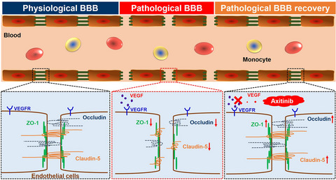
Lipoic acid protects the central nervous system by immunomodulation and antioxidation. In the periphery, LA prevents inflammatory cells from crossing the BBB by inhibiting the expression of LFA-1, ICAM-1, VLA-4, VCAM-1, and MMP-9 and protects brain endothelial cells. In the CNS, LA modulates autoimmunity by inhibiting the activity of T cells/microglia and decreasing the expression of TNF-α and IFN-γ, and LA reduces oxidative stress by neutralizing ROS and NO
LA inhibited the activity of immune cells and reduced inflammatory infiltration in the CNS. In terms of pathological evidence, five studies found that LA reduced CD3+/CD4+ T-cell infiltration in the brain and spinal cord, and three studies indicated reduced macrophage/microglial infiltration in EAE mice.81, 82 Above all, LA downregulated the expression of ICAM-1 and VCAM-1 in brain endothelial cells by colocalization analysis, contributing to the impairment of peripheral immune cell migration.81 For T cells, two studies revealed that LA increased Treg cell levels and decreased encephalitogenic T-cell levels, leading to a lower grade of the inflammatory response.72, 73 LA also inhibited T-cell activities by reducing the expression of MMP-9 in a dose-dependent manner to protect BBB integrity.69, 70 Furthermore, LA downregulated CD4 from the surface of Jurkat cells in a concentration-dependent manner. Interestingly, CD4 inhibitors reduced the severity of EAE symptoms in mice.83 These findings indicated that LA could effectively decrease the invasion of peripheral immune cells and the inflammatory response in the CNS.
NF-KB is a classical proinflammatory transcription factor existing in the cytoplasm, and it translocates to the nucleus and promotes the transcription of TNF-α and IL-6 during the oxidative stress.84 On the other hand, LA could directly inhibit the activity of NF-KB and its downstream protein expression.85 On the contrary, the enhanced antioxidative capability promoted by Nrf2 would also suppress the NF-KB activity. Besides, Nrf2 can inhibit the transcription of the various inflammatory mediators, including TNF-α and IL-6 in microglia and astrocytes.86, 87 In EAE mice, two studies indicated that LA decreased the expression of MMP-9 to protect the BBB from immune disruption.69, 70 Additionally, LA also decreased the expression of TNF-α, IFN-γ, and IL-4 to reduce the level of inflammation in EAE mice.
留言 (0)