The lateral posterior clock neurons (LPN) of Drosophila melanogaster express three neuropeptides and have multiple connections within the circadian clock network and beyond
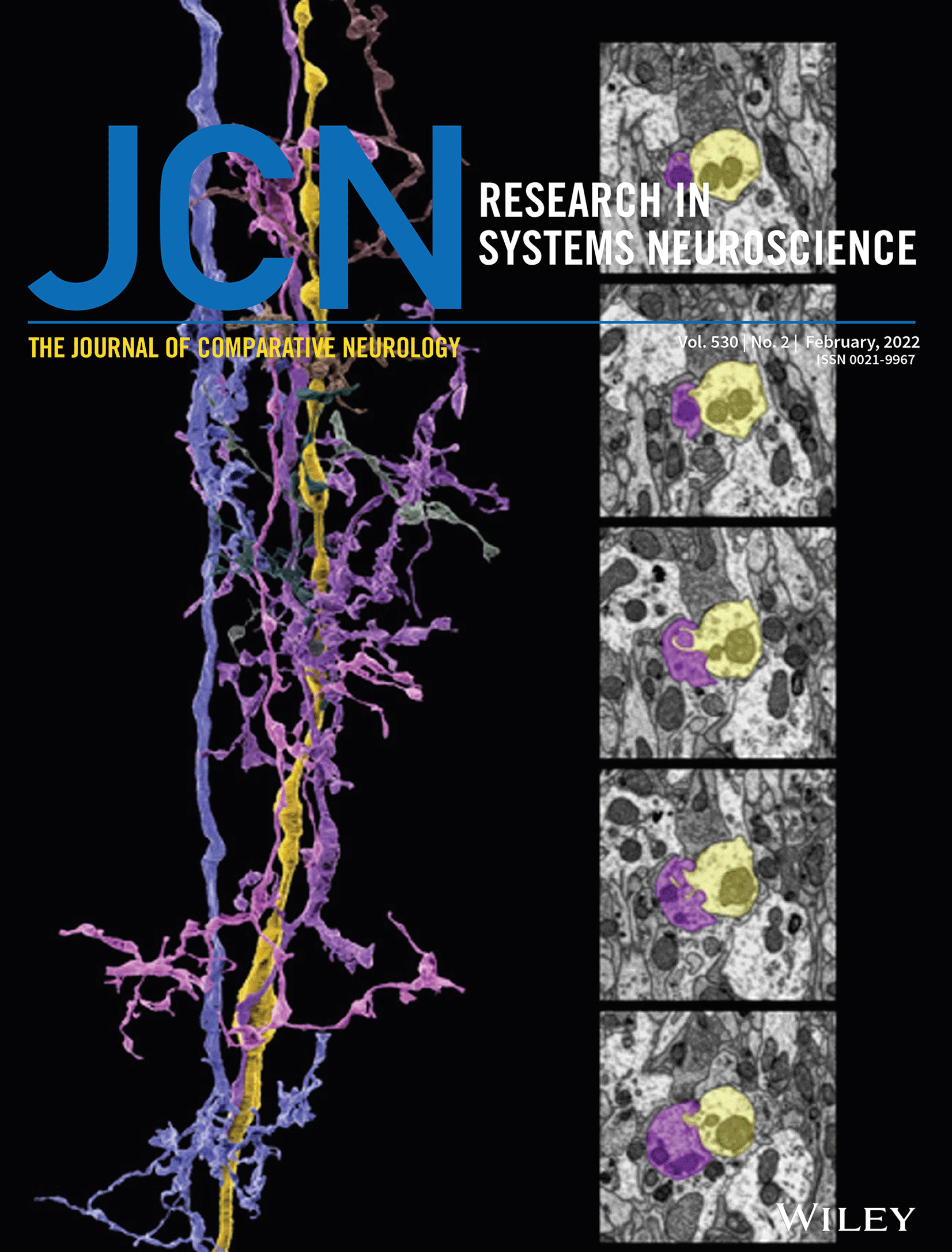
Drosophila’s lateral posterior neurons (LPNs) belong to a small group of circadian clock neurons that is so far not characterized in detail. Thanks to a new highly specific split-Gal4 line, here we describe LPNs’ morphology in fine detail, their synaptic connections, daily bimodal expression of neuropeptides, and propose a putative role of this cluster in controlling daily activity and sleep patterns. We found that the three LPNs are heterogeneous. Two of the neurons with similar morphology arborize in the superior medial and lateral protocerebrum and most likely promote sleep. One unique, possibly wakefulness-promoting, neuron with wider arborizations extends from the superior lateral protocerebrum toward the anterior optic tubercle. Both LPN types exhibit manifold connections with the other circadian clock neurons, especially with those that control the flies‘ morning and evening activity (M- and E-neurons, respectively). In addition, they form synaptic connections with neurons of the mushroom bodies, the fan-shaped body, and with many additional still unidentified neurons. We found that both LPN types rhythmically express three neuropeptides, Allostatin A, Allostatin C, and Diuretic Hormone 31 with maxima in the morning and the evening. The three LPN neuropeptides may, furthermore, signal to the insect hormonal center in the pars intercerebralis and contribute to rhythmic modulation of metabolism, feeding, and reproduction. We discuss our findings in the light of anatomical details gained by the recently published hemibrain of a single female fly on the electron microscopic level and of previous functional studies concerning the LPN.
This article is protected by copyright. All rights reserved


留言 (0)