
記住我


The intestine is a vast organ containing multiple cell types, including specialized epithelial, immune, and enteric neural cells with different subtypes. The latter form the enteric nervous system (ENS), a network of various subtypes of enteric neurons and enteric glia that differ in shape, function, and location, spanning the entire intestinal length.1, 2
Several types of enteric neurons (ENs) exist in the ENS, and the majority express choline acetyltransferase (ChAT).3 These cholinergic ENs are a prerequisite for developing a healthy gut4 and vital for intestinal motility5 and intestinal epithelium maintenance.6
Besides ENs, the ENS also harbors enteric glial cells (EGCs), a transcriptionally distinct type of glia in different shapes depending on their subtype,2 which expresses similar molecular markers as other glia populations like Schwann cells and oligodendrocytes.7 Similar to ENs, several distinct subtypes of EGC exist in the gut, yet over 95% of all EGCs express Sox10.8 Recent studies of us and others revealed that glia are involved in several gastrointestinal processes such as motility,9 intestinal inflammation,10 neurophysiological homeostasis, and barrier functions.2
Traditionally, sorting techniques, such as fluorescence-associated cell sorting (FACS), are utilized to generate data from specific cell types and gain a deeper understanding of those mechanisms. However, the common usage of antigens for sorting specific ENS cells exhibits several issues. Most conventional markers require extensive cell preparation or a fluorescent reporter signal. Additionally, these markers are often limited to niche subtypes, such as p75 for neural stem cells,11 that demand prior in vitro cultivation before the sorting procedure.12 This, together with the prerequisite sample preparation, mechanical and/or enzymatic disruption, and the sorting procedure itself introduces significant changes in cell composition (Figure 1A)13, 14 thereby complicating FACS. Moreover, neural cells possess a complex anatomical structure that is disrupted during sample preparation, causing loss of mRNA in cell processes,15 which limits RNA analysis. Another viable approach is the analysis of cell-specific transcriptomes via single-cell RNA-sequencing16 (scRNA-Seq; Figure 1A). This method is highly specific but includes a steep monetary cost, isolation-based handling bias, and its poor sequencing depth majorly limits comparison of different experimental conditions.17

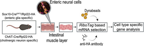
Comparative overview of RNA analysis methods. (A) Generation of specific cell population samples by fluorescence-based cell sorting and single-cell RNA-Seq analysis and their dis-/advantages. (B) Genetic overview of the Cre/Rpl22-HA construct (RiboTag). Excision of endogenous, loxP flanked Exon 4 of the Rpl22 gene by either tamoxifen-inducible Sox10iCreERT2 or ChATCre and subsequent expression of HA-tagged Rpl22 rRNA and tagged ribosomes. (C) RiboTag workflow: Schematic of a ribosome with Rpl22 subunit and incorporated HA-Tag carrying cell-specific mRNA, targeted cell types and their tissue location and workflow of sample preparation, IP and RiboTag approach advantages
As a solution to analyze tightly integrated and morphologically challenging cell types, Sanz et al. developed a method to extract mRNA from a specific cell type without prior cell separation—the RiboTag method15 wherein a transgenic Cre/loxP system is used to produce ribosomes with an Rpl22-haemagglutinin tag (RiboTag; Figure 1B) in the Cre-expressing cell type. This enables specific targeting and isolation of mRNA from in vivo cell populations by immune-precipitation (IP; Figure 1C). In the CNS, this method has been successfully applied for glia18, 19 and neurons.20 Meanwhile, the RiboTag approach was adapted to several cell types and peripheral tissues, for example, to analyze intestinal macrophages,21 by modifying the protocol toward unique cellular and extracellular matrix composition.17 Recently, first steps in utilizing the RiboTag approach for enteric neural cells have been undertaken.22-24 As the enteric nervous system has been shown to closely interact with the intestinal immune system and becomes transcriptionally active during inflammation, we utilized this method with either Sox10iCreERT2 or ChATCre mice to create the RiboTag in EGC or ChAT+ neurons, respectively (Figure 1B).
In this technical note, we provide a selective protocol including a comprehensive quantitative expression-based quality control in support of the RiboTag method as an appropriate tool to generate transcriptional in vivo snapshots of ENS cells without the need for elaborate sorting-based isolation.
留言 (0)