
記住我

Periprosthetic joint infection (PJI) is a rare but severe complication after total joint replacement (TJA)1, 2. It is a huge economic burden not only for the individual patient but also for the global healthcare system. For primary total joint arthroplasty, the incidence of infection ranges between 1 and 3%2. The current study indicated the infection ranges after revision total joint (hips and knees) arthroplasty 3% to 8%3. However, the diagnosis of infection is still the main challenge after TJA4. Besides, accurate differentiation between septic and aseptic failure is essential to determine treatment protocols5. There is currently lacking a single gold-standard test for diagnosing PJI. Based on the 2018 International Consensus Conference on Musculoskeletal infection, Surgeons rely on a combination of a series of laboratory tests in peripheral blood and synovial fluid, microbial culture, and histopathological examinations to diagnose PJI6. Recent studies have shown that potential novel markers that need to perform joint aspiration for the diagnosis of PJI, include synovial alpha-defensin, synovial leukocyte test strips, synovial C-reactive protein7-9. However, these potential synovial markers may increase the financial burden of patients. Moreover, surgeons are often facing a lack of synovial fluid or “dry tap”, and joint aspiration is an invasive procedure with an increased risk of deep infection. Improvements in diagnostic tools, mainly to obtain more accurate diagnostic information from blood tests, are necessary.
The blood test provides surgeons with important information on inflammatory markers due to its simplicity, accessibility, and short waiting time. Erythrocyte sedimentation rate (ESR) and C-reactive protein (CRP) are minor diagnostic criteria in 2018 ICM criteria. The reliability and usefulness of ESR and CRP for diagnosing PJI have been well studied. In a meta-analysis, the researchers noted the sensitivity and specificity were 0.860 (95% CI, 0.825–0.890) and 0.723 (95% CI, 0.704–0.742) for ESR, and for CRP were 0.869 (95% CI, 0.835–0.899) and 0.786 (95% CI, 0.769–0.803) according to the diagnostic criteria for ICM10. However, McArthur et al. indicated that both ESR and CRP are negative according to current diagnostic criteria, there are still cases involving false negatives for diagnosing PJI11. One study has also shown that the false-negative rate is 9.2 and 5.3% for ESR and CRP, respectively, combined ESR and CRP the false-negative rates up to 11.1%12. Controversy still exists due to their unclear thresholds and a negative serum ESR and CRP test result does not exclude the possibility of infection, so it is essential to obtain more accurate diagnostic information from the blood test results.
In addition to the classical inflammatory markers CRP and ESR, coagulation-related markers also showed high diagnostic efficacy in the diagnosis of PJI. Plasma fibrinogen and D-dimer have been studied by researchers for use to diagnose PJI13. Fibrinogen is a positive acute reactive protein produced by the liver and an essential component of the coagulation system. Li et al. demonstrated that the diagnostic efficacy of plasma fibrinogen was comparable to that of ESR and CRP14. Similarly, D-dimer is a fibrin degradation product, which is used as a screening tool for thrombosis and is also related to inflammation15. Some researchers noted that D-dimer showed better diagnostic efficacy than ESR and CRP levels5, 16. In addition to classic blood markers, several studies have documented that platelets and mean platelet volume (MPV) play an essential role in the inflammatory process13, 17, 18. Recent literature has shown that the combination of MPV/PC with CRP and ESR significantly improves the efficacy of diagnosing PJI19. Moreover, Huang et al. reported that peripheral blood dynamic ratio neutrophil/lymphocyte ratio (NLR) and MLR correlate with inflammatory body status20. It provides a new potential method for getting sufficient information for diagnosing PJI through a single blood test. However, more studies are needed to confirm the accuracy of ratio markers in the diagnosis of PJI and there is still a need to determine the diagnostic performance of the combination of coagulation-related markers with peripheral blood ratio markers.
In most institutions, it is usually possible to obtain a routine complete blood count by blood testing, including monocyte count, neutrophil count, lymphocyte count, platelet count, and MPV. Ratio valuables are also easy to calculate. Besides, a routine blood test can perform on all patients, particularly for those patients with “dry tap”. To the best of our knowledge, there are no studies that have evaluated the use of the combination of coagulation-related markers with peripheral blood dynamic ratio for the diagnosis of PJI.
Therefore, we hypothesized that peripheral blood indicators, coagulation-related markers combined with ratio markers maybe increase the accuracy of PJI diagnosis. The main purpose of the present study was to: (i) access the diagnostic value of ESR, CRP and coagulation-related markers; (ii) select useful ratio markers and examine the differences between bacterial species for ratio markers; and (iii) evaluate whether combined measurement provides surgeons additional information in order to diagnose PJI more accurately.
Materials and Methods Inclusion and Exclusion CriteriaThe inclusion criteria were: (i) patients diagnosed with chronic PJI or aseptic loosening (AL); (ii) patient underwent revision hip or knee arthroplasty; (iii) patients divided into two groups according to the 2014 MSIS criteria21; (iv) the major evaluation peripheral blood inflammatory biomarkers included CRP, ESR, plasma fibrinogen, d-dimer, WBC, PLR, PMR, NLR, MLR; and (v) this study is a retrospective study.
The exclusion criteria were: (i) acute PJI (defined as infection occurs within 3 months after the surgery); (ii) inflammation-related diseases include rheumatoid arthritis, systemic lupus erythematosus, and ankylosing spondylitis; (iii) revision due to periprosthetic fracture or dislocation; (iv) severe liver dysfunction or the presence of malignant tumors; and (v) insufficient serum marker data.
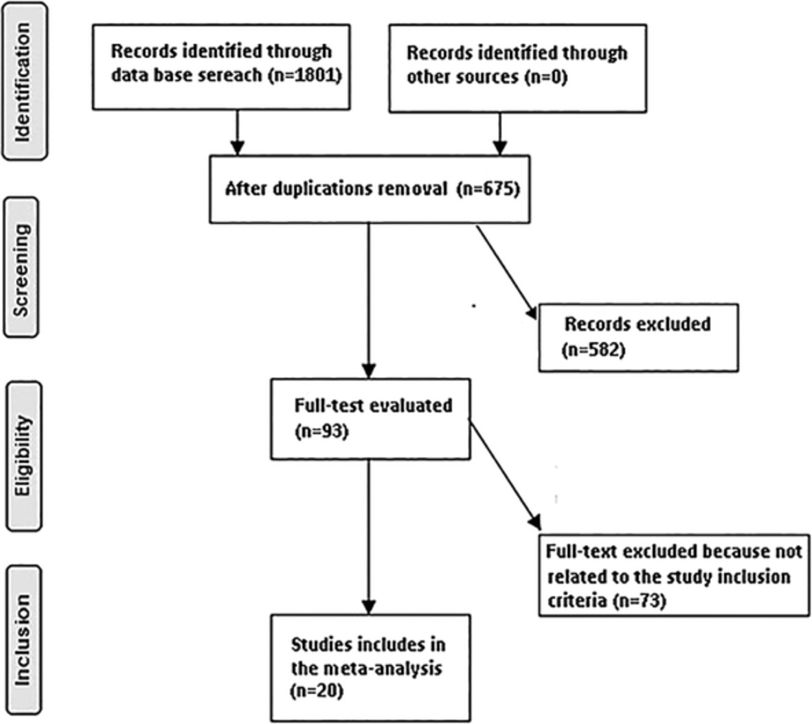
DemographicsOur institution's research ethics board approved this retrospective observational study. We enrolled patients who were managed with revision surgery after hip or knee arthroplasty from January 2017 to December 2018 at a single institution. Ultimately, 246 patients were included, of which 125 were diagnosed with PJI, and 121 were aseptic loosening (Fig. 1). The clinical records of the patient's information that included age, Body mass index (BMI), gender, surgery type (knee or hip), Charlson comorbidity index (CCI) were showed in Table 1.

Flowchart of included and excluded cases. PJI, periprosthetic joint infection; MSIS, Musculoskeletal Infection Society.
TABLE 1. Characteristics of the study group Variables PJI group (n = 125) Aseptic loosening group (n = 121) P value Age (year) 62.8 ± 12.7 61.2 ± 12.8 0.251 BMI (kg/m2) 25.7 ± 3.9 25.2 ± 3.7 0.417 Gender (%) 0.570 Female 72 (57.6%) 74 (61.2%) Male 53 (42.4%) 47 (38.8%) Joint <0.001 Knee 68 (54.4%) 31 (25.6%) Hip 57 (45.6%) 90 (74.4%) CCI (%) 0.266 <4 94 (72.8%) 106 (87.6%) ≥4 31 (27.2%) 15 (12.4%) BMI, body mass index; CCI, Charlson comorbidity index; PJI, periprosthetic joint infection. Laboratory Tests Blood Testing and DetailsAs part of our routine preoperative testing, blood samples are collected the morning after admission and then sent to the Medical Laboratory Center for testing. In addition to record ESR, CRP, fibrinogen, D-dimer, WBC as main peripheral markers; we also recorded lymphocyte count, neutrophil count, platelet count, and MPV to calculate PLR, PMR, NLR, MLR as adjunct ratio markers for diagnosing PJI. The CRP level was analyzed with a special protein analyzer PA-990 (Sysmex). The ESR was measured with an automated sed rate screener SRS 100/II (Greiner Bio-One). WBC count, platelet, and MPV were determined with an automated hematology analyzer, the Sysmex XN-20 modular system (Sysmex). Plasma fibrinogen and D-dimer were quantified with an STA R Max Evolution analyzer (Diagnostica Stago). A BACT/ ALERT 3D blood culture system (BioMerieux) was used for culture, and a matrix-assisted laser desorption ionization-time of flight mass spectrometry system, VITEK-MS (BioMerieux), was used for microorganism identification.
Other TestsOther tests include cultures, synovial leukocyte (WBC) counts, percentage of polymorphonuclear neutrophils (PMN%), and histological analysis. During the surgical procedure, about 3–5 samples (synovial fluid, deep tissue, and bone) were obtained for aerobic and anaerobic, and fungal cultures. The samples were sent to the medical laboratory center and each sample was inoculated for 14 days unless microorganisms were detected. Culture results were used for correlation analysis with ratio markers.
Statistical AnalysisAll the statistical analyses were performed with the statistical software packages R (http://www.R-project.org, The R Foundation). Categorical variables were expressed as frequencies and percentages, and continuous variables were expressed as mean ± standard deviation. A comparison of the clinical characteristics between the PJI group and AL group was performed using the independent t-test or the chi-square test. The diagnostic value of each marker for PJI assessment was determined by receiver operating characteristic ROC curve analysis. Curves are considered valuable when AUC ≥ 0.7. The Youden index was used to determine the optimal threshold for classical markers (fibrinogen, ESR, CRP, D-dimer) and ratio markers (PLR, PMR, NLR, MLR). Predictive models were utilized to screen the best combination of indicators. The statistical significance was defined as P < 0.05.
Results General ResultsDemographic information is shown in Table 1. This study included 246 patients, of whom 125 (50.8%) diagnosed with PJI and 121 (49.1%) with aseptic loosening. Of these, 99 patients underwent total knee revisions, and 147 patients undergone hip revisions. The mean ages of the PJI group and AL group were 62.8 ± 12.7 years and 61.2 ± 12.8 years (P = 0.251). There were no significant statistical differences between gender (P = 0.570), BMI (P = 0.471), and CCI (P = 0.266). The aseptic loosening group had more hip joints than the PJI group (90 [74.4%] vs 57 [45.6%]; P < 0.001).
Clinical Results Routine Blood TestsThe concentration of blood markers plasma fibrinogen, CRP, ESR, D-dimer, serum WBC, neutrophil count, monocyte count, the platelet count in the PJI group higher than the aseptic loosening group, except for lymphocyte count and mean platelet volume with statistically significant differences (P < 0.001) (Table 2).
TABLE 2. Distribution of blood markers in PJI and aseptic loosening group Markers PJI group Aseptic loosening group P value Mean ± SD Median (Min-Max) Mean ± SD Median (Min-Max) CRP (mg/L) 36.74 ± 48.05 19.22 (0.94–257.00) 5.77 ± 11.78 1.92 (0.50–81.00) <0.001 ESR (mm/h) 42.36 ± 28.33 36.00 (2.00–113.00)13.02 ± 11.42
10.00 (0.20–63.00) <0.001 D-dimer (μg/mL)1.80 ± 1.19
1.63 (0.15–6.99)1.33 ± 1.58
0.84 (0.16–11.36) <0.001 Plasma Fibrinogen (g/L) 5.74 ± 8.18 4.82 (2.40–95) 3.3 ± 0.79 3.1 (1.73–5.50) <0.001 WBC (109/L)6.88 ± 2.30
6.36 (2.16–18.00) 5.97 ± 1.42 5.75 (2.63–9.50) <0.001 Neutrophil count (109/L) 4.65 ± 2.05 4.24 (1.49–14.87)3.57 ± 1.09
3.33 (1.53–6.55) <0.001 Lymphocyte count (109/L) 1.54 ± 0.55 1.51 (0.20–2.86)1.77 ± 0.48
1.70 (0.75–2.85) <0.001 Monocyte count (109/L)0.47 ± 0.20
0.43 (0.20–1.66)0.37 ± 0.12
0.36 (0.17–0.74) <0.001 Platelet count (109/L) 293.43 ± 79.79 279 (106–511) 224.28 ± 52.19 22 (104–364) <0.001 Mean Platelet Volume (fl) 9.97 ± 1.01 9.90 (7.00–15.70) 10.67 ± 1.06 10.55 (8.30–14.20) <0.001 NLR3.52 ± 2.82
2.72 (1.06–25.47) 2.14 ± 0.81 2.01 (0.85–5.32) <0.001 MLR0.36 ± 0.22
0.28 (0.12–1.14)0.22 ± 0.08
0.21 (0.03–0.50) <0.001 PLR 236.60 ± 195.11 185.01 (108.12–1712.74) 135.19 ± 46.57 123.99 (55.08–290.47) <0.001 PMR29.73 ± 8.68
27.96 (10.00–56.09) 21.36 ± 6.01 21.68 (7.70–37.62) <0.001 CRP, C-reactive protein; ESR, erythrocyte sedimentation rate; MLR, monocyte to lymphocyte ratio; NLR, neutrophil to lymphocyte ratio; PLR, platelet to lymphocyte ratio; PMR, platelet to mean platelet volume ratio; WBC, white blood cell. ROC Curves Analysis for Various Laboratory MarkersUtilizing ROC curves to analyze the ability of major blood markers and adjunct ratio markers to diagnose PJI (Fig. 2). And calculating the AUC and the specificity, sensitivity, PPV, NPV of Blood markers. The AUC of plasma fibrinogen was 0.853 (95% CI, 0.805–0.901), followed by ESR 0.836 (95% CI, 0.785–0.887) and CRP 0.825 (95% CI, 0.773–0.878), respectively. While the AUCs of adjunct ratio markers were NLR 0.736 (95% CI, 0.674–0.798), MLR 0.733 (95% CI, 0.671–0.796), PLR 0.785 (95% CI, 0.729–0.840) and PMR 0.792 (95% CI, 0.736–0.847), which were more accurate than D-dimer 0.691 (95% CI, 0.624–0.758) and serum WBC 0.622 (95% CI, 0.552–0.692), respectively.

ROC curves for the diagnosis of PJI. (A) ROC curve analyses for classic inflammatory markers and coagulation-related markers. (B) ROC curve analyses for ratio markers. The black line depicts 50% sensitivity and specificity. NLR, neutrophil to lymphocyte ratio; PLR, platelet to lymphocyte ratio; PMR, platelet to mean platelet volume ratio; MLR, monocyte to lymphocyte ratio.
Comparison of Sensitivity and Specificity in PJI Group with AL GroupThe plasma fibrinogen optimal threshold (4.13 g/L) demonstrated a sensitivity and specificity of 75.20 and 86.78%, respectively. CRP and ESR had optimal cutoff of 7.34 mg/L (75.19% sensitivity, 84.30% specificity) and 26.0 mm/h. (69.60% sensitivity, 88.43% specificity), respectively. PLR had an optimal threshold of 129.33 (83.20 and 57.02%). The optimal threshold of MLR at 0.26 (60.00% sensitivity, 81.82% specificity) and the optimal threshold of PMR at 23.42 (79.80% sensitivity, 68.33% specificity). The WBC and D-dimer were unable to demonstrate comparable diagnostic efficacy to the adjunct ratio indicators (Table 3).
TABLE 3. ROC analysis for main and ratio markers Test AUC (95%CI) Best threshold Specificity Sensitivity Postive-pv Negative-pv Plasma Fibrinogen (g/L) 0.8531 (0.8051–0.9011) 4.1300 0.8678 0.7520 0.8545 0.7721 CRP (mg/L) 0.8252 (0.7725–0.8780) 7.3350 0.8430 0.7519 0.8319 0.7669 ESR (mm/h) 0.8359 (0.7852–0.8865) 26.00 0.8843 0.6960 0.8614 0.7379 D-dimer (μg/mL) 0.6913 (0.6243–0.7584) 1.2350 0.6860 0.6560 0.6833 0.6587 Serum WBC (109/L) 0.6219 (0.5521–0.6917) 7.5500 0.8760 0.3280 0.7321 0.5579 NLR 0.7358 (0.6739–0.7977) 2.4185 0.7107 0.6720 0.7059 0.6772 MLR 0.7332 (0.6707–0.7957) 0.2550 0.8182 0.6000 0.7732 0.6644 PLR 0.7846 (0.7292–0.8399) 129.3250 0.5702 0.8320 0.6667 0.7667 PMR 0.7918 (0.7362–0.8473) 23.4150 0.6833 0.7980 0.7164 0.7387 MLR, monocyte to lymphocyte ratio; NLR, neutrophil to lymphocyte ratio; PLR, platelet to lymphocyte ratio; PMR, platelet to mean platelet volume ratio. Distribution of Ratio Markers between Bacterial SpeciesDifferences between Gram-positive and Gram-negative bacteria in inflammatory predictive markers were presented in Table 4. This result highlights no significant differences in peripheral blood markers between microorganisms.
TABLE 4. Comparison between Gram positive PJI and Gram-negative PJI Types Gram-positive (N = 91) Gram-negative (N = 9) P value PMR 29.21 ± 8.52 27.21 ± 11.39 0.227 PLR 235.09 ± 191.99 318.98 ± 320.20 0.608 MLR 0.35 ± 0.19 0.47 ± 0.36 0.529 NLR 3.34 ± 1.86 6.54 ± 8.03 0.468 MLR, monocyte to lymphocyte ratio; NLR, neutrophil to lymphocyte ratio; PLR, platelet to lymphocyte ratio; PMR, platelet to mean platelet volume ratio. Results of the Prediction ModelThe combination of PMR and MLR was selected as the best ratio indicators combination among all ratio markers based on the prediction model calculation (Table 5, Fig. 3). The result of single main variables combined with both PMR and MLR are as follows, plasma fibrinogen achieved the highest AUC of 0.923 (95% CI, 0.891–0.951), which resulted in a sensitivity, specificity, PPV, and NPV of 0.864, 0.841, 0.850, and 0.856, respectively. Similarly, PMR, MLR combined with ESR and CRP, the AUC raised to 0.91 (95% CI, 0.880–0.946), 0.899 (95% CI, 0.861–0.931), respectively. When multiple variables including all peripheral main markers (plasma fibrinogen, CRP, ESR, D-dimer, serum WBC) and ratio markers (NLR, PLR, MLR, PMR) were placed in the prediction model, the best combination was plasma fibrinogen, ESR, and CRP, PMR, and MLR. The new combination results in good sensitivity, specificity, PPV, and NPV are 0.824, 0.892, 0.888, and 0.823, respectively (Fig. 4). Compared with the use of plasma fibrinogen, CRP, and ESR alone, the combined use of ratio markers shows a good diagnostic value (P < 0.001). A combination of single ratio markers with single main markers shown in Table S1.
TABLE 5. Comparison of ROC curves between single and multiple combined markers Main markers vs combination with ratio markers AUC (95%CI) Specificity sensitivity PPV NPV P-value Plasma fibrinogen vs plasma fibrinogen + PMR + MLR 0.9233 (0.8909–0.9508) 0.8417 0.8640 0.8504 0.8559 <0.001 CRP vs CRP + PMR + MLR 0.8994 (0.8606–0.9307) 0.8167 0.8400 0.8268 0.8305 0.0072 ESR vs ESR+ PMR + MLR 0.9159 (0.8804–0.9456) 0.8250 0.8640 0.8372 0.8534 <0.001 Plasma fibrinogen + ESR + CRP vs plasma fibrinogen + ESR + CRP+ PMR + MLR 0.9269 (0.8909–0.9547) 0.8917 0.8240 0.8879 0.8295 <0.001 AUC, area under the curve.
Comparison of receiver operator curves between single and multiple combined markers. ROC for fibrinogen and combined MLR and PLR predicting PJI. (B) ROC for ESR and combined MLR and PLR predicting PJI. (C) ROC for CRP and combined MLR and PLR predicting PJI. Fbg, fibrinogen; PMR, platelet to mean platelet volume ratio; MLR, monocyte to lymphocyte ratio.

ROC curves of best blood marker combination by the prediction model. Fbg, fibrinogen; PMR, platelet to mean platelet volume ratio; MLR, monocyte to lymphocyte ratio.
DiscussionPJI is a devastating complication after TJA. It is estimated that the rate of PJI approximately 1% to 3% of patients who undergo primary TJA, and it will increase the mental and financial burden of patients2, 4, 22, 23. Recent studies indicate that novel tests such as leukocyte esterase (LE), alpha-defensin, and next-generation genome sequencing show very promising diagnostic efficacy, but these tests are expensive, require specialized equipment, and cannot be implemented in all institutions7-9, 24, 25. Also, these tests cannot be used as a single gold standard for the diagnosis of PJI, and according to the 2018 MSIS criteria combination of various clinical examination approaches is still recommended. Therefore, identifying reliable and accurate potential markers for the diagnosis of PJI is the key to the preoperative diagnosis and the development of an appropriate treatment plan. The primary purpose of this study was to determine the diagnostic value of ratio markers and evaluate the performance of the combined diagnosis of PJI with classical inflammatory and coagulation-related markers.
Diagnostic Value of ESR, CRP, and Coagulation-related MarkersThe results of the classical inflammation-related markers ESR, CRP, and coagulation-related marker plasma fibrinogen were consistent with previous studies, demonstrating promising diagnostic efficacy (AUCs > 0.8)14. However, the findings of the D-dimer of the current study do not support the previous research that D-dimer outperformed both the ESR and the CRP5, 16. There were two possible explanations, one is D-dimer concentration is different in races, another is the previous study use the plasma as the tested sample, but in our study, we use serum sample as a tested sample, which may result in different levels of D-dimer. In our study, the diagnostic power of the inflammation ratio indicators (AUCs > 0.7) was significantly better than the D-dimer (AUC: 0.691). As we find, one of the factors influencing peripheral blood parameters is bacterial species.
Differences of Ratio Markers Between Bacterial SpeciesIn a previous study, Abe et al.26 demonstrated that blood inflammation markers CRP and IL-6 had significantly higher Gram-negative bacteremia than Gram-positive bacteremia. These findings suggest that differences in host response and virulence mechanisms to different pathogenic microorganisms should be taken into account when using peripheral blood ratio indicators. The findings of Kalbian et al. confirm the prevailing view that Gram-negative PJI is associated with poorer overall results than Gram-positive PJI27. Therefore, determining the diagnostic efficacy of markers is very important, and different bacterial species may influence other indicators. However, our study indicated no significant differences in peripheral blood markers between microorganisms. As a ratio indicator, they may not be significantly affected by Gram-positive versus Gram-negative bacteria according to the present result. Another possible explanation for this might be that delayed infections may be associated with low-virulen
留言 (0)