
記住我
The circle of Willis (CoW) is an anastomotic arterial network located on the base of the brain. It functions to prevent cerebral and cerebellar ischaemia by maintaining tissue perfusion given an impaired or decreased blood flow through one or more of its component vessels (Hartkamp et al., 1999; Kapoor et al., 2008).
The CoW is divided into two sections. The anterior communicating artery (AComA) and A1 segments of the anterior cerebral arteries (A1) form the anterior half of the circle (Gray, 2016; Moore et al., 2014). The posterior communicating arteries (PComA) and P1 segments of the posterior cerebral arteries (P1) form the posterior half of the circle (Gray, 2016; Moore et al., 2014). The arteries create a symmetrical polygonal-shaped connection between the internal carotid and vertebrobasilar systems.
Four criteria are classically used to define ‘normal’ (non-variant) anatomy of the CoW: (1) all segments (AComA, A1s, PComAs and P1s) are present (De Silva et al., 2011; Eftekhar et al., 2006; Kapoor et al., 2008; Klimek-Piotrowska et al., 2015; Vasović et al., 2013), (2) all segments arise from their natural origins (De Silva et al., 2011; Kapoor et al., 2008; Klimek-Piotrowska et al., 2015), (3) no accessory arteries are present (De Silva et al., 2011; Kapoor et al., 2008; Klimek-Piotrowska et al., 2015; Vasović et al., 2013), and (4) all segments have an external diameter of >1 mm (De Silva et al., 2011; Kapoor et al., 2008; Klimek-Piotrowska et al., 2013, 2015; Vasović et al., 2013).
The prevalence of anatomical variation of the CoW in the neurologically healthy human population is estimated to be 68.22 ± 14.32% (Jones et al., 2020). For this review, anatomical variation was defined using two criteria: (1) the variation is embryologically derived, and (2) the variation does not demonstrate the potential to directly progress to a pathological consequence. Commonly recorded variation types include hypoplasia (Cilliers et al., 2018; De Silva et al., 2011; Eftekhar et al., 2006), absence (Hafez et al., 2007; Klimek-Piotrowska et al., 2013; Li et al., 2020), and duplication (Iqbal, 2013; Klimek-Piotrowska et al., 2015). The most common variant segment is the posterior communicating artery (Eftekhar et al., 2006; Hindenes et al., 2020; Klimek-Piotrowska et al., 2015).
An awareness of the anatomical variations of the CoW is important for clinical practice (Jones et al., 2020; Raikos & Smith, 2015). Circle variation is associated with an increased risk of a number of cerebrovascular diseases (Henry et al., 2015; Oumer et al., 2021; Ryan et al., 2015; Stojanović et al., 2019), and affects patient response to therapeutic intervention (Leng et al., 2016; Wufuer et al., 2017). It has implications for preoperative planning and is important in selecting the most appropriate method of cerebral protection (Papantchev et al., 2013). Anatomical variation influences a range of intraoperative factors which determine patient outcome and increases the risk of misinterpretation and surgical error. Despite this, there is lack of consensus in the literature on a system that comprehensively documents and classifies such variations.
Three classification systems are commonly used to categorise anatomical variations of the CoW in humans. The Riggs classification system (Riggs & Rupp, 1963) contains 21 variations. Circle order in this classification has no relation to anatomy, only segment hypoplasia is included, and complete anatomical descriptions are not provided. No single diagram of the included circles is available. In contrast, the Lazorthes classification system (Lazorthes et al., 1979) contains 22 variations. Here, only segment hypoplasia is included, and circle order has no relation to anatomy. The illustration of segment hypoplasia is inconsistent, creating ambiguity regarding the anatomy of variation numbers five, seven, eight, 13 and 17 (Lazorthes et al., 1979). Finally, the Krabbe-Hartkamp classification system (Krabbe-Hartkamp et al., 1998) contains 18 variations. Complete circle anatomy is not shown, and artery hypoplasia and absence are not differentiated.
Four studies (De Silva et al., 2011; Eftekhar et al., 2006; Klimek-Piotrowska et al., 2015; Vasović et al., 2013) assert that Riggs (Riggs & Rupp, 1963) and Lazorthes (Lazorthes et al., 1979) describe, and include in their classification systems, a variant circle with a hypoplastic AComA, a unilateral hypoplastic A1 and an ipsilateral hypoplastic PComA. However, both classification systems (Lazorthes et al., 1979; Riggs & Rupp, 1963) contain a circle with a unilateral hypoplastic A1 and an ipsilateral hypoplastic PComA without a hypoplastic AComA. As such, Riggs (Riggs & Rupp, 1963) and Lazorthes (Lazorthes et al., 1979) classification systems have been incorrectly used throughout the literature. Interestingly, no study in this review, nor those aforementioned, has recorded a circle with a hypoplastic AComA, a unilateral hypoplastic A1 and an ipsilateral hypoplastic PComA. In view of this lack of consensus in the literature, a recent review (Jones et al., 2020) on the prevalence of anatomical variation of the CoW recommended the development of a new, comprehensive classification system.
This article, therefore, presents a systematic review of published empirical research on anatomical variation of the CoW in humans, performed with the following aims: (1) to identify and catalogue the described anatomical variations of the CoW in humans, and (2) to characterise the described variants to produce a new, comprehensive classification system.
2 METHODSThe review was conducted according to the recommendations set out in the Preferred Reporting Items for Systematic Reviews and Meta-Analyses (PRISMA) statement (Shamseer et al., 2015).
A systematic search of the published peer-reviewed literature was conducted on the Ovid Medline (1946 to May 27, 2020), Ovid Embase (1974 to May 27, 2020), Web of Science Core Collection (all years 1900–2020), Scopus and The Cochrane Library databases on 27 May 2020. The search strategy was made up of two sections. The first section contained alternative terms for ‘circle of Willis’ and its component arteries. The second section contained synonyms of the term ‘variation’ used in anatomical description. The strategy was adapted to each database to increase search sensitivity (Table 1). No limitations on language or publication format were applied.
TABLE 1. The search strategies Database Search strategy Ovid Medline (1946 to May 27, 2020)(exp “Circle of Willis”/OR circle of Willis OR cerebral arterial circle OR circulus arteriosus cerebri OR circulus arteriosus Willis* OR circulus Willis* OR Willis* circle OR Willis* polygon OR exp Anterior Cerebral Artery/OR anterior cerebral arter* OR arteria cerebri anterior OR exp PCA/OR posterior cerebral arter* OR arteria cerebri posterior OR anterior communicating arter* OR arteria communicans anterior OR posterior communicating arter* OR arteria communicans posterior)
AND
(exp Anatomic Variation/OR varia* OR anomal* OR abnormal* OR atypical OR incomplete OR unusual)
Ovid Embase (1974 to May 27, 2020)(exp brain circulus arteriosus/OR circle of Willis OR cerebral arterial circle OR circulus arteriosus cerebri OR circulus arteriosus Willis* OR circulus Willis* OR Willis* polygon OR Willis* circle OR exp anterior cerebral artery/ OR anterior cerebral arter* OR arteria cerebri anterior OR exp PCA/OR posterior cerebral arter* OR arteria cerebri posterior OR exp anterior communicating artery/ OR anterior communicating arter* OR arteria communicans anterior OR exp posterior communicating artery/OR posterior communicating arter* OR arteria communicans posterior)
AND
(exp anatomic variation/OR varia* OR anomal* OR abnormal* OR atypical OR incomplete OR unusual)
Web of Science Core Collection (all years 1900–2020)(TS = (“circle of Willis” OR “cerebral arterial circle” OR “circulus arteriosus cerebri” OR “circulus arteriosus Willis*” OR “circulus Willis*” OR “Willis* polygon” OR “Willis* circle” OR “anterior cerebral arter*” OR “arteria cerebri anterior” OR “posterior cerebral arter*” OR “arteria cerebri posterior” OR “anterior communicating arter*” OR “arteria communicans anterior” OR “posterior communicating arter*”))
AND
(TS = (“varia*” OR “abnormal*” OR “anomal*” OR “atypical” OR “incomplete” OR “unusual”))
Scopus(TITLE-ABS-KEY(“circle of Willis” OR “cerebral arterial circle” OR “circulus arteriosus cerebri” OR “circulus arteriosus Willis*” OR “circulus Willis*” OR “Willis* polygon” OR “Willis* circle” OR “anterior cerebral arter*” OR “arteria cerebri anterior” OR “posterior cerebral arter*” OR “arteria cerebri posterior” OR “anterior communicating arter*” OR “arteria communicans anterior” OR “posterior communicating arter*” OR “arteria communicans posterior”))
AND
(TITLE-ABS-KEY(“varia*” OR “abnormal*” OR “anomal*” OR “atypical” OR “incomplete” OR “unusual”))
The Cochrane Library(exp Circle of Willis/ OR circle of Willis OR cerebral arterial circle OR circulus arteriosus Willis* OR circulus Willis* OR Willis* circle OR exp Anterior Cerebral Artery/OR anterior cerebral arter* OR arteria cerebri anterior OR exp PCA/OR posterior cerebral arter* OR anterior communicating arter* OR posterior communicating arter*)
AND
(exp Anatomic Variation/OR varia* OR anomal* OR abnormal* OR atypical OR incomplete OR unusual)
2.1 Screening processFollowing deduplication, studies underwent a two-phase screening process against inclusion and exclusion criteria (Table 2). In phase one, titles and abstracts were screened against criteria In1, In5, and Ex1-5. Criteria In2, 3, 4 and 6 were not applied as titles and abstracts provided insufficient evidence to inform decisions on their fulfilment. For studies meeting all inclusion criteria and no exclusion criteria, full texts were sought, and English translations obtained when freely available.
TABLE 2. The inclusion and exclusion criteria Inclusion criteria Exclusion criteria - In1. The title or abstract mention an anatomical variation of the CoW - In2. The anatomy of the CoW is described or illustrated in its entirety - In3. Any intracerebral arterial variation is exclusive to the AComA, A1s, PComAs and/or P1s - In4. The anatomical descriptions and/or illustrations are clear and specific - In5. The study is primary research - In6. The study is available in the English language - Ex1. The study exclusively: Ex1a. Identifies a variant anatomical course of one or more arteries of the CoW Ex1b. Investigates the haemodynamics of one or more arteries of a variant CoW Ex1c. Investigates the calibre of one or more arteries of the CoW - Ex2. A foetal study population without a non-foetal human control - Ex3. A non-human study population without a non-foetal human control - Ex4. The study uses cerebral vascular models - Ex5. The study is published as a conference abstractIn phase two, full texts were screened against all inclusion and exclusion criteria. To meet criterion In2, studies were required to describe or illustrate the anatomy of the AComA, A1 segments, PComAs and P1 segments of the variant circle. To meet criterion In4, studies were required to describe or illustrate the type of variation identified. Angiographic studies which did not differentiate between artery hypoplasia and absence were excluded. This included the Krabbe-Hartkamp classification (Krabbe-Hartkamp et al., 1998). Studies meeting one or more exclusion criteria were removed, while studies with one or more circles meeting all inclusion criteria were included in the review. Two independent reviewers screened two random samples of 15 studies using the inclusion and exclusion criteria. No difference to the original study selection was found.
2.2 Data extractionData were extracted on the background of the study (first author and publication year), its characteristics (location, design, population, modality, and definition of hypoplasia), and its description of variant circles (variant segment(s), variation type(s), position of the variation, and the number of different variant circles).
2.3 Quality assessmentA bespoke quality assessment tool was created, adapting the modified Anatomical Quality Assessment Tool (Henry et al., 2017) and Critical Appraisal Skills Programme Checklist for Case-Control and Cohort Studies (Critical Appraisal Skills Programme, 2018a, 2018b) criteria. Case-control, cohort and cross-sectional studies underwent quality assessment. Case studies and case series were not assessed as the tool was incompatible with the study types. The quality assessment tool is shown in Table S1.
Seven study domains were scored: clearly defined and focused aim(s), appropriate study design, representative population characteristics, reproducible methodology, clarity of descriptive anatomy, accuracy in reporting of results, and acknowledgement of limitations. A quality threshold score was subjectively determined. Studies scoring ≥14 out of 25 were considered high quality, while studies scoring <14 out of 25 were considered low quality. Studies were not excluded from the review on the basis of quality score.
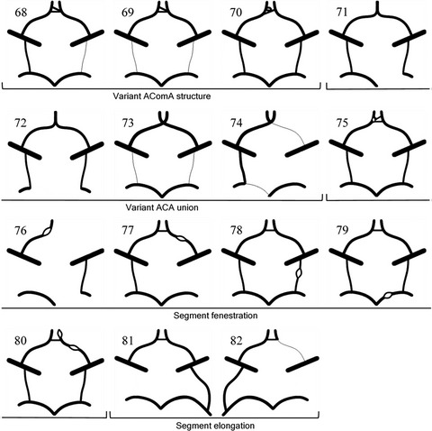
2.4 Data synthesisThe circles were reconstructed digitally using Paint 3D (Microsoft Corporation, 6.2003.4017.0). A1 and P1 segments were drawn using the 4-point curve tool at 11 px (pixel). The AComA and PComAs were drawn using the 2-point line tool at 7 px. Internal carotid and middle cerebral arteries, drawn using the 2-point line tool at 18 px, were included for anatomical completeness. Artery hypoplasia was shown by line width conversion to 1 px.
The left-right orientation of variant segments is rarely reported within the literature. For standardisation, the most anterior variation was drawn on the right side of the circle.
The circles were grouped according to variation type. Within each group, circles were subcategorised according to the number or type of variant segment. A coding system was created to simplify anatomical description of the circles.
3 RESULTSA summary of the study selection process is shown in Figure 1. The search identified 14,437 studies. After removal of 8538 duplicates, 5899 studies underwent title and abstract screening, of which 5375 did not meet the assessed criteria. Of the resulting 524 studies, 29 full texts were unobtainable. The remaining 495 studies underwent full-text screening. A total of 42 studies were included in the review.

A flow diagram summarising the study selection process
3.1 Study characteristics and resultsA summary of the study characteristics and results is shown in Table 3. Studies were published between 1903 and 2020. Five different study designs were used: cross-sectional (n = 19), case report (n = 17), case-control (n = 2), case series (n = 2) and prospective cohort (n = 2). The study locations spanned Asia (n = 15), Europe (n = 14), North America (n = 9), Africa (n = 3) and South America (n = 1). A range of investigative modalities were used: cadaveric dissection (n = 22), computed tomography angiography (n = 4), digital subtraction angiography (n = 3) and magnetic resonance angiography with or without 3D time-of-flight capability (n = 15). Three studies used two investigative modalities and one study used cerebral cast angiography.
TABLE 3. A table showing the characteristics and results of studies included in the review Study code Study Study location Study design (study population [n]) Study modality Definition of artery hypoplasia Distinct variants (n) Quality assessment score (n/25) S1 Al-Hussain et al. (2001) Jordan CSS (50) CD <1 mm 4 11 S2 Benson et al. (1986) Canada CR (1) CD N/A 1 N/A S3 Cilliers et al. (2018) South Africa CSS (59) CD <1 mm 10 15 S4 De Silva et al. (2011) Sri Lanka CSS (225) CD <1 mm 15 18 S5 Ding et al. (2019) China CR (1) CD N/A 1 N/A S6 Drummond et al. (2006) USA CR (1) TOF MRA N/A 1 N/A S7 Drummond et al. (2012) USA CR (1) TOF MRA N/A 1 N/A S8 Eftekhar et al. (2006) Iran CSS (102)a CD <1 mm 10 15 S9 Giglio et al. (2010) Italy CR (1) MRA N/A 1 N/A S10 Gurdal et al. (2004) Turkey CR (2) CD N/A 2 N/A S11 Hafez et al. (2007) Egypt CSS (130) 3D TOF MRA and CD <1 mm 7 11 S12 Hashemi et al. (2013) Iran CSS (200) CD <1 mm 9 12 S13 He et al. (2016) China PCS (102) CTA <1 mm 1 15 S14 Howe (1903) USA CR (1) CD N/A 1 N/A S15 Howie (1959) USA CCS (256) CD N/A 10 4 S16 Ibrahim et al. (2017) Sudan CCS (146) 3D TOF MRA N/A 5 10 S17 Iqbal (2013) India CSS (50) CD <1 mm (AComA and PComA <0.5 mm) 5 13 S18 Jensen et al. (2017) USA CR (1) DSA N/A 1 N/A S19 Karatas et al. (2016) Turkey CSS (100) CD N/A 2 14 S20 Klimek-Piotrowska et al. (2013) Poland CSS (250) CTA N/A 3 14 S21 Klimek-Piotrowska et al. (2015) Poland CSS (100) CD <1 mm 30 17 S22 Li et al. (2020) China CSS (819) 3D TOF MRA and DSA <1 mm 7 16 S23 Loh and Sharma (2010) Singapore CR (1) TOF MRA N/A 1 N/A S24 Malamateniou et al. (2009) UK CSS (103) 3D TOF MRA N/A 3 18 S25 Manninen et al. (2009) Finland CSS (92) Cerebral cast angiography N/A 1 19 S26 Matsuda et al. (2017) Japan CR (1) MRA N/A 1 N/A S27 McCullough (1962) USA CS (77) CD N/A 4 N/A S28 Ozturk et al. (2008) Turkey CR (1) CD N/A 1 N/A S29 Papantchev et al. (2007) Bulgaria CSS (112) CD <1 mm 3 12 S30 Papantchev et al. (2013) Bulgaria CSS (500) CD and CTA <1 mm 6 16 S31 Riggs and Rupp (1963) USA CSS (994) CD N/A 20 7 S32 Sabau et al. (2012) Romania CR (1) 3D TOF MRA N/A 1 N/A S33 Saikia et al. (2014) India CSS (70) TOF MRA <1 mm 2 14 S34 Saphir (1935) USA CR (3) CD N/A 3 N/A S35 Sonobe et al. (2019) Japan CR (1) MRA N/A 1 N/A S36 Sonobe et al. (2020) Japan CR (1) MRA N/A 1 N/A S37 Stefani et al. (2013)
留言 (0)