Brain TDP‐43 pathology in corticobasal degeneration: topographical correlation with neuronal loss
Aims
Neuronal and glial inclusions comprising transactive response DNA-binding protein of 43 kDa (TDP-43) have been identified in the brains of patients with corticobasal degeneration (CBD), and a possible correlation between the presence of these inclusions and clinical phenotypes has been speculated. However, the significance of TDP-43 pathology in the pathomechanism of CBD has remained unclear. Here we investigated the topographical relationship between TDP-43 inclusions and neuronal loss in CBD.
Methods
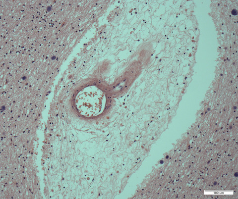
We estimated semi-quantitatively neuronal loss and TDP-43 pathology in the form of neuronal cytoplasmic inclusions (NCIs), astrocytic inclusions (AIs), oligodendroglial cytoplasmic inclusions (GCIs), and dystrophic neurites in 22 CNS regions in 10 patients with CBD. Then, the degree of correlation between the severity of neuronal loss and the quantity of each type of TDP-43 inclusion was assessed. We also investigated tau pathology in a similar manner.
Results
TDP-43 pathology was evident in 9 patients. The putamen and globus pallidus were the regions most frequently affected (80%). NCIs were the most prominent form, and their quantity was significantly correlated with the severity of neuronal loss in more than half of the regions examined. The quantities of TDP-43 NCIs and tau NCIs were correlated in only a few regions. The number of regions where the quantities of TDP-43 AIs and GCIs were correlated with the severity of neuronal loss was apparently small in comparison with that of NCIs.
Conclusions
TDP-43 alterations in neurons, not closely associated with tau pathology, may be involved in the pathomechanism underlying neuronal loss in CBD.


留言 (0)