
記住我

In this study, we mainly aimed to (1) determine the HR & LD protocol with MBIR and/or DLR to achieve the lowest radiation dose and the similar low-frequency noise to that using the routine protocol in the phantom study and (2) assess validity of this HR & LD protocol based on image quality and radiologists’ acceptance using the routine protocol as reference in the clinical pilot study.
Our institutional review board approved this clinical study, and we obtained written informed consent from all patients.
Phantom studyPhantomsWe assessed spatial resolution by the task-based modulation transfer function (TTF) using a quality assurance phantom (TOS phantom; Canon Medical Systems, Tochigi, Japan) that included inserts of various materials to provide different levels of image contrast (air, -1000 HU; polypropylene, -105 HU; water, 0 HU; acrylic, 120 HU; Delrin, 340 HU; and Teflon, 940 HU). We focused on the acrylic insert of the lowest positive contrast, almost equivalent to the contrast between soft tissue and fat attenuations, for oncologic follow-up by CE-APCT. To assess image noise characteristics by the noise power spectrum (NPS), we utilized an original abdomen phantom comprising an elliptical cylinder (33-cm longest diameter, 22-cm shortest diameter) made of epoxy-based and polyurethane resin (Kyoto Kagaku, Kyoto, Japan) (Fig. 1) [16].
Fig. 1
Axial CT images of the 2 phantoms used to assess a the task-based modulation transfer function (TTF), which included cylinder inserts of various materials that offered different image contrasts (left to right: Delrin, 340 HU; acrylic, 120 HU; air, -1000 HU; polypropylene, -105 HU; and Teflon, 940 HU), and b the noise power spectrum (NPS), made primarily of epoxy-based and polyurethane resin
CT image acquisition and reconstructionWe performed helical scanning of the 2 phantoms with a UHRCT scanner (Aquilion Precision, Canon Medical Systems) using automatic exposure control (AEC) and parameters for our routine and HR & LD protocols, which are summarized in Table 1. Specifically, we used 5 dose settings at AEC noise indices of 20, 25, 30, 35, and 40 HU for the HR & LD protocol at 100 kV. To assess radiation exposure, we reviewed the volume CT dose index (CTDIvol) for each protocol recorded as a dose report.
Table 1 Parameters for CT scanning and reconstructionWe reconstructed the phantom images using a standard kernel (FC03) and an HIR algorithm (Adaptive Iterative Dose Reconstruction [AIDR] 3D Standard, Canon Medical Systems) for those acquired with the routine protocol and using an MBIR algorithm (Forward-projected model-based Iterative ReconSTruction [FIRST] Body Standard, Canon Medical Systems) and a DLR algorithm (Advanced intelligent clear-IQ engine [AiCE] Body Standard, Canon Medical Systems) for those acquired with the HR & LD protocol. Table 1 summarizes other reconstruction parameters.
For the DLR, as shown in Fig. 2, standard-dose images by HIR as low-quality input data and high-dose images by advanced MBIR with much more iterations than MBIR as targeting high-quality data were used as training pairs, and in the training process, statistical features that differentiate signals from the noise and artifacts could be “learned” and then “updated” in the deep convolutional neural network for use in future reconstructions [6, 17]. This training process had been performed in advance as a black box by the manufacturer. Because these ideal MBIR images were used to train the network, DLR yielded comparable or superior image quality to that of MBIR in a shorter processing time than that of MBIR [6].
Fig. 2
Flowcharts of the training and reconstruction process in deep learning reconstruction (DLR). a In the training process, given standard-dose images by hybrid iterative reconstruction (HIR) as low-quality input data and high-dose images by advanced model-based iterative reconstruction (MBIR) with much more iterations than MBIR as targeting high-quality data as training pairs, the deep convolutional neural network (DCNN) is updated to minimize the difference between DCNN output and the target for future reconstructions. This process has been performed in advance as a black box by the manufacturer. b In the reconstruction process, the DCNN is validated for clinical image processing to generate final high-quality images from input images by HIR
Image quality assessmentWe analyzed each set of phantom images using the appropriate software (CT measure version 0.98, http://www.jsct-tech.org/; Excel 2016, Microsoft). On an axial image of the acrylic insert in the quality assurance phantom, we radially acquired and averaged profile curves crossing the circular edge to obtain its edge-spread function. TTF was calculated by Fourier transformation using line-spread function obtained by differentiating the edge-spread function to assess the intermediate-contrast in-plane spatial resolution with non-linear algorithms, such as HIR, MBIR, and DLR, at various noise levels. Our method to determine NPS in the epoxy-based resin part is described elsewhere [8, 16]. Using routine images reconstructed by HIR for reference, we then compared TTF and NPS between the HR & LD images with the 5 dose settings at noise indices of 20, 25, 30, 35, and 40 HU reconstructed by MBIR and DLR. Ultimately, we determined the noise index setting for the AEC in the HR & LD protocol by MBIR and/or DLR to minimize the radiation dose while preserving or lessening noise at lower frequencies compared with the noise obtained using the routine protocol.
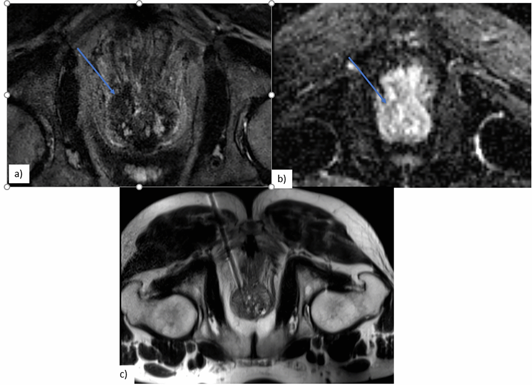
Clinical pilot studySubjectsFrom March 11 through March 29, 2019, we prospectively enrolled 41 consecutive adult patients with mild to moderate renal impairment (i.e., estimated glomerular filtration rate: 30 to 59 mL/min/1.73m2) from whom we obtained written informed consent and who underwent CE-APCT using an HR & LD protocol with the UHRCT scanner for oncologic follow-up. Exclusion criteria were: inadequate CT image acquisition and history of surgical operation of the liver and/or intrapelvic organs, which precluded our image quality assessment described below. Actually, 5 patients were excluded due to insufficient scan coverage (n = 3) and history of total hysterectomy (n = 2). Thus, we finally included 36 consecutive patients (24 men, 12 women; mean age, 75 ± 9 years; range, 48 to 93 years; mean body weight [BW], 57.1 ± 11.7 kg; range, 39 to 86 kg; mean body mass index [BMI], 22.5 ± 3.5 kg/m2; range, 15 to 29 kg/m2) in the present study. Using the routine protocol as reference, we compared the quality of the CT images reconstructed by MBIR and DLR. In the HR & LD protocol, we set the noise index at 35 HU based on the results of the aforementioned phantom study, as described in the “Results” section, and reduced the iodine load by 40% of that with our routine protocol.
CT image acquisition and reconstructionPatients underwent helical acquisition of CE-APCT with the UHRCT scanner using parameters summarized in Table 1. All patients received non-ionic iodinated CM (Iopamiron 300; Bayer HealthCare, Osaka, Japan) at a concentration of 300 mgI/mL. A total dose of 312 mgI/kg of BW was administered over 45 s via the right antecubital vein using a 22-gauge plastic intravenous catheter with a power injector (Dual Shot-type GX 7; Nemoto Kyorindo, Tokyo, Japan), and scanning began at 120 s following the start of CM administration. To assess radiation exposure, we reviewed the CTDIvol and dose-length product (DLP) recorded as a dose report and then calculated the estimated effective dose as the DLP multiplied by a k factor for the abdomen and pelvis of 0.015 mSv mGy−1 cm−1 [18] for each patient. Thus, we calculated the mean CTDIvol, DLP, and estimated effective dose for the HR & LD protocol. As in the phantom study, we used both the MBIR and DLR algorithms to reconstruct the CE-APCT images acquired for each patient (Table 1).
Quantitative assessment of image qualityOn the CT images reconstructed by both MBIR and DLR and displayed on a commercially available workstation (Ziostation Version 2.4; Ziosoft, Inc., Tokyo, Japan), 3 radiology technologists, by consensus, employed a copy-and-paste function to place 3 circular regions of interest (ROIs) in the hepatic parenchyma, carefully avoiding large vessels and any areas of focal changes in attenuation, and prominent artifacts. In a similar manner, they placed a circular ROI in the upper abdominal subcutaneous fat at the same level, in the major psoas muscle at the level of the aortic bifurcation, avoiding any macroscopic fat infiltration, in the urinary bladder and the prostate (for men) or uterus (for women), avoiding any areas of focal change in attenuation and prominent artifacts, and in the lower abdominal subcutaneous fat at the same level. Thus, they measured the CT number and its standard deviation (SD) value within these anatomies in each patient. We calculated the mean SD value in all patients as objective noise in the hepatic parenchyma, upper abdominal subcutaneous fat, major psoas muscle, urinary bladder, and lower abdominal subcutaneous fat. We also calculated the contrast-to-noise ratio (CNR) of the liver and pelvis using the following equations: CNR of the liver = (mean CT number of the hepatic parenchyma – CT number of the major psoas muscle)/noise in the upper abdominal subcutaneous fat, and CNR of the pelvis = (CT number of the prostate or uterus – CT number in the urinary bladder) / noise in the lower abdominal subcutaneous fat.
Qualitative assessment of image qualityOn the workstation, 2 independent board-certified radiologists with 10 and 11 years’ clinical experience who were blinded to patient demographics and CT parameters used a 5-point scale to grade the quality of CE-APCT images reconstructed by both MBIR and DLR. Five points represented much better quality compared with the reference; 4 points, better quality; 3 points, comparable quality; 2 points, worse quality; and one point, much worse quality. Referencing routine CE-APCT images of other 101 consecutive adult patients (52 men, 49 women; mean age, 65 ± 17 years; range, 29 to 92 years; mean BW, 57.3 ± 14.4 kg; range, 32 to 117 kg; mean BMI, 21.9 ± 4.5 kg/m2; range, 12 to 36 kg/m2) with normal renal function (i.e., estimated glomerular filtration rate: ≥ 60 mL/min/1.73m2) imaged by UHRCT using the routine scan and reconstruction protocol (Table 1) and our routine iodine load (520 mgI/kg of BW) from January 1 through March 1, 2019, the reviewers considered the general acceptability of the image with regard to overall diagnostic confidence and both image appearance and image texture in the liver and intrapelvic organs (prostate or uterus and urinary bladder) as well as image noise, as described by Laurent and colleagues [7]. The HR & LD images reconstructed by both MBIR and DLR were presented in random order on a preset soft tissue window (window width, 370 HU; window level, 40 HU).
Statistical analysisResults were expressed as mean ± SD for continuous variables. Statistical analysis was performed using commercially available statistical software (SPSS for Windows, Version 23.0, IBM SPSS, Armonk, NY). Objective noise and CNR were compared between MBIR and DLR using paired t-test, and subjective image quality grades were compared using Wilcoxon signed-rank test. BW and BMI were compared between the study and reference patient groups using unpaired t-test. A P value below 0.05 was considered to indicate significant difference. Inter-reviewer agreement was estimated using weighted kappa statistics.
留言 (0)