
記住我
This study was conducted on subjects with neuromuscular diseases with pneumonia admitted to a university hospital from August 2015 to July 2016. Pneumonia was diagnosed based on clinical symptoms, and laboratory and radiologic findings. Evidence of cardinal infection such as fever or chills and leukocytosis, signs or symptoms localized to the respiratory system (cough, increased sputum production, and increased respiratory rates), and a new or changed infiltrate on simple radiography were analyzed. All patients enrolled in the study received intravenous antibiotics therapy, for treatment of pneumonia, and the study was conducted during treatment.
The following patients were excluded: those with a risk for barotrauma and a history of pneumothorax or lung bullae; those in whom PCF could not be checked because of severe bulbar dysfunction; those who need continuous ventilator support, and could not perform the test; and those who showed unstable vital signs. We did not exclude pediatric patients; however, we excluded patients with impaired cognitive function.
Study protocol We applied the MI-E to patients according to protocol of our institute, based on cumulative experiences and other previous research. During the application, patients were in an upright or semi-upright sitting position. Settings of the MI-E were +40 cmH2O and -40 cmH2O pressure with insufflation and exsufflation times of 2–3 and 1–2 seconds, respectively [13]. Patients underwent a total of five cycles, with a pause of 20–30 seconds between each cycle to prevent hyperventilation [14]. PCF without assistance and assistive PCF (APCF) were evaluated sequentially as follow: PCF and APCF before MI-E application, PCF and APCF 15 minutes after MI-E, and PCF and APCF 45 minutes after MI-E. Non-assisted PCF was measured by asking the patient to inhale maximum amount of air unassisted, before coughing as hard as possible. APCF was measured by first asking the patient to inhale maximum amount of air unassisted, breathing in additional amount of air through a mask by using a manual resuscitator bag, and then an assistant applied a strong push to the patient’s abdomen while the patient was coughing [10]. Each process was repeated at least three times, and maximum value was selected from measurements (Fig. 1). Protocol was approved by the Institutional Review Board of Gangnam Severance Hospital (No. 3-2015-0149).Statistical analysis was performed using IBM SPSS statistics version 20 (IBM, Armonk, NY, USA). Repeatedmeasures analysis of variance was used to compare values of PCF and APCF at each time, with Bonferroni correction for post hoc analysis.
RESULTS A total of 27 patients (11 with Duchenne muscular dystrophy, 10 with progressive muscular dystrophy, 3 with ALS, 2 with spinal muscular atrophy, and 1 with limbgirdle muscular dystrophy) were included. Mean age was 30.0±13.3 years (range, 12–66 years), and 21 of them (77.8%) were men. All patients had a functional disability and could not walk without support. Twenty-four of them were using non-invasive ventilator support, and none of them was using invasive ventilator support (Table 1). In baseline evaluation before application of MI-E, initial PCF value was 92.6±57.1 L/min and the APCF value was 170.7±66.0 L/min. At 15 minutes after application of MI-E, measured PCF was 100.4±57.2 L/min and the APCF was 179.3±12.4 L/min. At 45 minutes later, PCF was 100.7±60.9 L/min and the APCF was 184.1±75.5 L/min. The increase in PCF and that in APCF compared with their baseline values, were both statistically significant (p=0.001). Comparing baseline PCF and APCF with measured value after 15 minutes, a statistically significant difference was found for both parameters (p=0.03, p=0.016). However, comparing PCF and APCF values measured after 15 minutes with those measured after 45 minutes, peak flow was increased but the difference between time points was not statistically significant (Table 2, Fig. 1). DISCUSSION This study demonstrated that PCF and APCF increase for a certain period, after use of MI-E in patients with neuromuscular disease and pneumonia. According to previous studies, compared with unassisted coughing and other standard cough augmentation techniques (physiotherapy-assisted, non-invasive ventilator-assisted, and exsufflation-assisted coughing), the MI-E significantly increased PCF in patients with neuromuscular disease [10-13]. However, although existing studies have demonstrated increase in PCF during or immediately after use of MI-E, none of the studies showed whether the effects persist for a certain period. This study is the first to demonstrate increased PCF even after a certain period after application.Maintaining increased cough flow is more critical in patients treated for current respiratory infection, because all of them have coughing disability and trouble with eliminating secretion. Nonetheless, whereas many previous studies usually excluded patients with pneumonia, we chose patients treated for pneumonia.
Many patients with neuromuscular disease, have respiratory muscle weakness that leads to declined lung compliance. Consistent increase in PCF may be explained as increase in compliance of the lung and chest wall. According to previous studies, lung compliance and the capacity for breathing increased for 3 hours, upon MI-E application in patients with kyphoscoliosis or ALS [15,16]. We can assume that the forced vital capacity and PCF, are increased temporarily by increased lung compliance. Furthermore, increased cough flow may be due to temporary conditioning, and strengthening effects on the respiratory muscle of the stretching exercise [17]. Stretching of respiratory muscles facilitates actin-myosin interaction, and strengthens respiratory muscles. Reduced airway resistance after removal of respiratory secretions, is also a probable mechanism, and has been proven in many previous studies.This study has limitations. We assumed that the 15 minutes time point reflects the short-term effect of MI-E and 45 minutes reflect the lasting effect. However, 45 minutes is not sufficient time to reflect lasting effect of treatment. Maximum insufflation capacity, maximal expiratory pressure, and maximal inspiratory pressure were checked only at baseline, and only PCF and APCF were checked during follow-up. It is also a limitation that the mechanism of increasing PCF, was not definitively explained. A longer-termed and detailed evaluation is needed to explain lasting effect of this treatment. Severity, type, and disease course of pneumonia, as well as patient status at the moment of evaluation during the hospital stay, were not considered in detail, all of which could have an effect on response to treatment with MI-E.
In addition, the training effect may also be considered. It would be difficult to explain the consistent increase in PCF after an intervention, as all subjects were repeatedly evaluated in PCF and APCF consecutively during followup periods also. Therefore, the training effect should be considered, as repetitive instructions also increase subjects’ compliance with using MI-E.
As this was not a blinded or randomized control study, we could not conclude whether increased PCF affect the course of pneumonia. The effect of increased PCF in patients with neuromuscular disease and pneumonia could have been more definitely evaluated, if another group such as patients with pneumonia not using MI-E or patients without pneumonia was compared. Further case–control studies involving a large group of subjects and more detailed evaluation, are needed to explain the accurate mechanism for these effects.
In conclusion, increased ability to expectorate sputum was confirmed after using MI-E, which persisted for at least 45 minutes after therapy. MI-E is a useful device for patients with neuromuscular disease and pneumonia, not only as the best way to manage secretion, but also as an intervention to maintaining coughing ability.
CONFLICTS OF INTERESTNo potential conflict of interest relevant to this article was reported.
Fig. 1.(A) PCF and (B) APCF before and after mechanical in-exsufflator application. Each graph shows the mean (dot) and standard error (bar). PCF, peak cough flow; APCF, assisted peak cough flow.
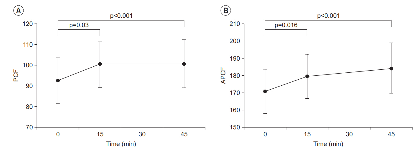 Table 1.
Baseline characteristics of patients
Characteristic Value Age (yr) 30.0±13.3 Sex Male 21 Female 6 Use of non-invasive ventilator 24 (82.8) Respiratory parameters FVC (mL) 652.4±296.6 FVC (% of predicted value) 16.6±8.9 MIP (cmH2O) 16.6±9.4 MIP (% of predicted value) 19.7±13.4 MEP (cmH2O) 19.2±12.7 MEP (% of predicted value) 16.3±13.5 Table 2.Measured PCF according to time
Before 15 min 45 min p-valuea) Post-hoc analysis (p-value) Before vs. 15 min 15 min vs. 45 min PCF (L/min) 92.6±11.0 100.4±11.0 100.7±11.7 <0.001 0.030 >0.999 APCF (L/min) 170.7±12.7 179.3±12.9 184.1±14.5 <0.001 0.016 0.158 REFERENCES 1. Bach JR, Ishikawa Y, Kim H. Prevention of pulmonary morbidity for patients with Duchenne muscular dystrophy. Chest 1997;112:1024–8.
留言 (0)