
記住我


Pancreatic cancer (PC) has been becoming the second cause of cancer death in the western world,1, 2 even though its incidence is relatively low (7th or 8th depending on the studies). Moreover, the incidence of PC is rising sharply in some countries with unclear reasons. The incidence of PC has increased with a speed of 3% each year since 1980 in France,3 and similar increasing trends were observed in the United States during the past four decades.4 The prognosis of PC remains poor with a 5-year survival rate for all stages combined around 8%.1, 2, 5 It was thus still a big challenge for us to cure patients with PC and improve their survival time.
Surgical resection is the only curative measure for patients with PC at present. However, 80% of PC patients may be unresectable at the time of diagnosis, because PC is generally aggressive and evolves rapidly which causes local-regional advances and metastases. That is to say surgical resection can only be performed in patients with locally confined tumors, without locoregional vascular invasion or distance metastases. Although there have been significant advances in patient selection, surgical techniques, and the perioperative care of patients with PC in the past 25 years, the 5-year survival rate is still poor (between 7 and 24%) in the patients who undergo surgery.6 One of the important factors influencing survival is whether an R0 resection was achieved.7, 8 The majority of PC resections were margin involvement (R1) resection, which significantly affects the prognosis of PC patients.9, 10 As a consequence, a strategy of adjuvant therapy following surgery is conventionally applied for PC patients. The Gastrointestinal Tumor Study Group (GITSG) trial reported that the median survival for the adjuvant therapy group was significantly longer than the no adjuvant therapy arm (20 vs 11 months).11 A major problem of adjuvant therapy is that a large percent of patients fail to receive a designed adjuvant therapy because of postoperative complications, delayed surgical recovery, patient refusal, comorbidity, or early disease recurrence.12-14
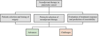
The neoadjuvant (preoperative) therapy is a promising strategy to overcome the challenges in the treatment of PC. Advantages of this approach included the following several aspects: (1) provide more patients with the chance of receiving surgery; (2) increase the likelihood of achieving R0 margins; (3) decrease the difficulty of the surgical procedure; (4) improve access of most patients to chemotherapy; and (5) clear circling tumor cells in blood and lymph vessels to avoid postoperative recurrence. Although a lot of efforts have been made in the field of neoadjuvant therapy for PC, several questions remain controversial as follows: which types of patients should be given to neoadjuvant therapy? If selected to neoadjuvant therapy, what therapeutic regimens should be used? If targeted drugs and immunotherapy are feasible as neoadjuvant therapy? After the start of neoadjuvant therapy, how should we evaluate the therapeutic response and determine the timing of surgery? We thus write this article to review the latest evidence about neoadjuvant therapy of PC and discuss the most recent advances, aiming to give a picture of the trends in this field.
2 PATIENTS SELECTION AND TIMING OF SURGERYThe classification of PC is various across different cancer institutions. Based on the status of resectability which is defined by multiple disciplinary teams, the National Comprehensive Cancer Network (NCCN) guideline classifies PC as resectable disease, borderline resectable disease, locally advanced disease, and metastatic disease.15 The American Society of Clinical Oncology (ASCO) published three clinical practice guidelines for the management of PC in 201616-18 and classified PC as three groups, that is, potentially curable PC, locally advanced PC (LAPC), and metastatic PC, which was mainly based on the tumor biological behaviors. Different classification criteria may cause inconsistent recommendations on the timing of surgery and selection of patients for neoadjuvant therapy. As a consequence, we can find a big controversy exists on the indications of neoadjuvant treatment across different guidelines.
The ASCO guideline refers to neoadjuvant therapy for any of the following19: radiographic findings suspicious but not diagnostic for extrapancreatic disease, a performance status or comorbidity profile not currently appropriate (but potentially reversible) for a major abdominal operation, a radiographic interface between primary tumor and mesenteric vasculature on cross-sectional imaging that does not meet appropriate criteria for primary resection or a CA 19-9 level (in absence of jaundice) suggestive of disseminated disease. In addition, the ASCO guideline also suggested that neoadjuvant therapy should be offered as an alternative treatment strategy for any patient who meets the surgical criteria. The strengths of these recommendations are high. Thus, the ASCO guideline is lenient on the selection criteria for neoadjuvant therapy patients. As long as the possibility of surgical resection exists, patients can receive neoadjuvant therapy before surgery. The NCCN guideline is constantly changing and updating the criteria for patient selection of neoadjuvant therapy. For resectable diseases, the 2018 NCCN guideline version 220 suggested that neoadjuvant therapy should be considered in high-risk patients (ie, patients having the following features: imaging findings, very highly elevated CA 19-9, large primary tumors, large regional lymph nodes, excessive weight loss, extreme pain), and it is preferred to be carried out in clinical trials. However, in the 2019 NCCN guideline version 1,21 it is modified as "Consider neoadjuvant therapy, particularly in high-risk patients,” indicating that the indications of neoadjuvant therapy for resectable diseases have been expanded. For borderline resectable diseases, the NCCN guidelines published after 2016 no longer recommend the strategy of either upfront surgery or neoadjuvant therapy followed by resection, but neoadjuvant therapy as the mandatory treatment. Indeed, the ASCO guidelines directly recommend that preoperative therapy be administered to patients with indications of extrapancreatic disease and those with poor performance status, whereas the NCCN guidelines do not directly address these clinical variables, and instead produced different algorithms for each class of pancreatic patients based on tumor anatomy.
Despite rapid advances in surgical techniques, postoperative local recurrence and non-R0 resection of PC are still common.22-24 The margin status is directly associated with the prognosis of patients. Chandrasegaram et al performed a systematic review and revealed a reduction in the risk of death by comparing the R0 resection group to the R1 group.9 Similarly, another systematic review by Kim et al. evaluated the relationship between resection margin distance and mortality risk, showing that the risk of death at group of 0, 0-1, and >1 mm resection margins decreased successively.10 Obviously, the status of the resection margin is one of the important outcomes in PC surgery, and the achievement of R0 resection is crucial to improve the prognosis of patients with PC. Many failures of R0 resection are due to the lack of preoperative accurate assessment and tumor staging, in addition to the inexperienced operative skills of surgeons. It is very limited to rely on CT to predict R0 resection. Actually, R0 resection may still not achieve during the surgical process in the patients who are predicted as resectable by CT. Therefore, whether there is a strong likelihood of achieving R0 margins can be one of the criteria for the selection of patients with neoadjuvant therapy. All patients with a high likelihood of R1 margins should be treated with neoadjuvant therapy because neoadjuvant therapy can significantly improve the rate of R0 resection.25, 26 In addition, PC is now considered a systemic disease, and long-term survival is associated with a variety of factors. In the selection of surgical timing for PC patients, we should consider the condition of patients comprehensively. Any factor that may indicate poor prognosis but that neoadjuvant therapy can improve its influence can become an indicator of preoperative therapy. In short, all patients who would benefit from neoadjuvant therapy should undergo neoadjuvant therapy.
The concept of neoadjuvant therapy is not applicable to the unresectable patients, and a more appropriate description should be conversion therapy. The NCCN guidelines have clearly stated that patients with locally advanced disease should be given priority to conversion therapy. However, the ASCO guideline thinks that it is difficult to convert unresectable tumors into resectable tumors with the use of systematic treatment, and this guideline thus focuses mainly on local control of tumors and improvement of quality of patients’ life. There are many new findings at present on the surgery conversion effectiveness of conversion therapy and its significance for patients with locally advanced disease. A prospective study conducted by Hackert et al27 showed that in patients with LAPC, the successful surgery conversion rate after neoadjuvant therapy was 50.8%. In addition, surgical resection after neoadjuvant therapy can improve the median overall survival (OS) of patients (15.3 months after resection vs 8.5 months after exploration alone, P < 0.0001). The study from Gemenetzis et al28 also showed that among the locally advanced patients who underwent surgical exploration after neoadjuvant therapy, 72% patients achieved resection of the primary tumor, and the patients who underwent surgical resection had significantly higher median OS compared with those who did not (35.3 vs 16.3 months, P < 0.001). The researches are still going on, and the surgical opportunities of these previously unresectable PC patients and their survival outcomes have been increasing, all of which benefit from the application of neoadjuvant therapy strategies. Obviously, patients with locally advanced diseases are also one of the appropriate populations for neoadjuvant therapy (conversion therapy).
It can be briefly summarized that the indications of neoadjuvant therapy have continuously expanded in the PC populations based on the findings of great benefit from neoadjuvant therapy. Neoadjuvant therapy has been widely tested in both resectable and unresectable diseases, but challenges still exist. For example, the inconsistent classifications and recommendations regarding neoadjuvant therapy among different guidelines limited its clinical application in PC.
3 PROTOCOLS SELECTION OF NEOADJUVANT THERAPYCurrently, most of the available evidence for the recommendations of the neoadjuvant chemotherapy regimen is the experience of a single institution, which lacks the support of high-quality and reliable evidence. There are a variety of chemotherapy regimens, but the types of regimens evaluated in a single study are often limited, making it difficult to draw comprehensive conclusions. In addition, there is great heterogeneity in the design of chemotherapy regimens across different studies, which makes the results from different studies uncomparable, then leads to the inability to draw reliable conclusions. In the process of history, the popular chemotherapy regimens also changed over time. The early regimen was based on fluorouracil (5-fu), and the regimens after 1997 were mainly based on gemcitabine. In 2011 and 2013, multiagent chemotherapy regimens Nab-Paclitaxel plus Gemcitabine and FOLFIRINOX (a combination of 5-FU, folinic acid, ilinotecan, and oxaliplatin) were demonstrated to be more effective to improve OS and progression-free survival (PFS) in the metastatic setting compared to gemcitabine alone. The introduction of FOLFIRINOX and Nab-Paclitaxel plus Gemcitabine regimens for metastatic disease has signed a watershed in the neoadjuvant treatment too. In recent years, FOLFIRINOX and Nab-Paclitaxel plus Gemcitabine regiments have been attracting more and more attention in the field of neoadjuvant therapy.
FOLFIRINOX for neoadjuvant therapy was the most commonly tested regimen in current researches. Several studies have shown the satisfying effectiveness of the FOLFIRINOX regimen. The first study was conducted by Hosein et al in 2012. Hosein et al showed us a good response rate of FOLFIRINOX based on a small sample (18 patients) study.29 Subsequently, a high surgical resection rate (including R0 resection) of FOLFIRINOX as neoadjuvant therapy was highlighted by several studies. Mahaseth et al30 evaluated the efficacy of FOLFIRINOX on 60 patients diagnosed with either borderline resectable, locally advanced, and metastatic cancer. They found a radiologically detectable response in 30% of patients, and resection was performed in 42% (6/14) of patients with an R0 rate of 83% (5/6). An observational cohort by Marthey et al31 evaluated the tolerability and efficacy of this regimen in LAPC and metastatic disease. They reported tumor resection for 28 (36%) patients with 25 (89%) having complete resection. They also reported a manageable toxicity profile with only 6% of treatment withdrawal because of tolerability problems. Sadot32 reported an experience of Memorial Sloan Kettering Cancer Center between 2010 and 2013. A total of 101 patients were included in this study and 31 patients (31%) finally underwent resection with an R0 resection rate of 55%. A large study of 575 patients selected for surgical exploration following neoadjuvant therapy was conducted by Hackert et al.27 FOLFIRINOX was administered to 125 individuals. A successful resection was achieved in 292 patients (50.8%), with the highest resection rate and R0 rate found in the FOLFIRINOX group (60.8% [76/125] vs 48% [216/450] achieved with other treatments, P = 0.011 and 40.8% [31/76] vs 30.1% [65/216], P = 0.048, respectively). In addition, although some studies have reported that neoadjuvant therapy with FOLFIRINOX regimen has a positive impact on patient survival outcomes, most studies have not reported it due to difficulties in evaluating survival outcomes. In recent years, some researchers confirmed the effectiveness of FOLFIRINOX to achieve a potentially curative resection and encouraging progress in prognosis by using the method of systematic review.33-36
The researches of Nab-Paclitaxel plus Gemcitabine regimen using for neoadjuvant therapy were very limited. However, we can still see its great potential from the few studies that have been done. Ielpo et al37 compared the results of neoadjuvant therapy with Nab-Paclitaxel plus Gemcitabine versus surgery first in 81 patients with either resectable disease or borderline resectable PC (BRPC). A resection rate of 68.8% was detected in the neoadjuvant therapy group. The median OS of patients who underwent surgery was higher in the neoadjuvant therapy group (30.6 months) compared with those in the surgery first group (22.1 months) (P = 0.04). Similarly, another study from Itchins et al38 showed that the overall resection rate was 79% with the R0 rate being 75% for the administration of Nab-Paclitaxel plus Gemcitabine in patients with resectable and BRPC. A similar survival rate was found between Nab-Paclitaxel plus Gemcitabine and gemcitabine alone or FOLFIRINOX group (mOS 23.0 vs 29.0 vs 25.9 months, respectively, log-ranked P = 0.92).
The FOLFIRINOX and Nab-Paclitaxel plus Gemcitabine regimens have become two popular regimens for neoadjuvant therapy due to its favorable treatment response (Table 1). The NCCN guideline15 recommends these two regimens as first-line regimens for neoadjuvant treatment in PC. However, the ASCO guidelines19 state that no data are available to support one regimen over another. In short, the current evidence of neoadjuvant chemotherapy is insufficient to rank the priority of different regimens. Although the FOLFIRINOX and Nab-Paclitaxel plus Gemcitabine regimens have shown tremendous potential, some thorny issues remain such as the management of toxicities. The studies evaluating the toxicities of the FOLFIRINOX and Nab-Paclitaxel plus Gemcitabine regimens for neoadjuvant therapy are lacking at present, but limited data showed that toxicities are common in patients receiving these regimens, with 64% grade 3 or higher adverse events for FOLFIRINOX40 and 52% for Nab-Paclitaxel plus Gemcitabine.49 The control of toxicities in a low level is more important when administering the regimens before surgery than after surgery, because severe toxicities may induce a delay of radical surgery. To reduce toxicities, one of the reasonable solutions is the adjustment of dose. For example, modified FOLFIRINOX by reducing the initial dose or dose intensity can significantly lower the frequency and severity of toxicities but did not influence the activity compared to the standard FOLFIRINOX.50, 51
TABLE 1. Studies on supporting FOLFIRINOX and Nab-Paclitaxel + Gemcitabine as neoadjuvant regimens in pancreatic cancer Outcomes Study Country Study design Patients R0 resection rates Survival Adverse events FOLFIRINOX Okada39 Japan Multicenter pilot trial 10 BRPC, 5 in the 4-cycle modified FOLFIRINOX arm, 5 in the 8-cycle arm4-cycle arm: 4 resections (3 R0)
8-cycle arm: 3 resections (2 R0)
– any grade events (100%), grade 3–4 events (80%) Katz40 USA Prospective multicenter single-arm trial 22 BRPC with modified FOLFIRINOX followed by capecitabine-based chemoradiation 15 resections (14 R0, 1 R1) Median OS: 21.7 months (95% CI, 15.7 to not reached) grade 3 or higher events (64%) Lakatos41 Hungary Prospective single center study 32 LAPC with modified FOLFIRINOX 2 resections (all R0)Median time to disease progression: 148 days (IQR: 58-228)
Median time to death: 312 days (IQR: 225-450)
The most frequent grade 3–4 events: nausea (18.8%), fatigue (12.5%), diarrhea (12.5%) de Marsh42 USA Proof-of-concept pilot study 21 RPC with modified FOLFIRINOX 17 resections (16 R0)Median OS: 34 months (95% CI 12.3-57.6)
PFS rate at 24 months: 0.27 (95% CI 0.10-0.47)
The most common adverse events: diarrhea,anorexia, and fatigue. Grade 4 events (rare) Murphy25 USA Single-arm phase 2 clinical trial 48 BRPC with FOLFIRINOX followed by individualized chemoradiotherapy 32 resections (31 R0)Median OS: 37.7 months (95% CI, 19.4 to not reached)
Median PFS: 14.7 months (95% CI, 10.5 to not reached)
grade 3 or greater events (19%), the most common severe events: diarrhea (n = 5), neutropenia (n = 2), and peripheral neuropathy (n = 2) Tran43 USA Single-institution phase 2 trial 25 BRPC with FOLFIRINOX and intensity modulated radiation therapy 13 resections (all R0)Median OS: 24.4 months (95% CI, 12.6-40.0)
Median PFS: 13.1 months (95% CI, 7.3-24.7)
grade3-4 events: neutropenia (40%), nausea and vomiting (28%), diarrhea(16%), fatigue (12%) Murphy44 USA Single-arm phase 2 clinical trial 49 LAPC with FOLFIRINOX and losartan 34 resections (30 R0)Median OS: 31.4 months (95% CI, 18.1-38.5)
Median PFS: 17.5 months (95% CI: 13.9-22.7)
grade 3 or greater events (51%), the most common severe events: neutropenia (n = 7), thrombocytopenia (n = 7), diarrhea(n = 7), nausea/vomiting (n = 4) Petrelli33 Italy Systematic review and meta-analysis 253 BRPC and URPC with FOLFIRINOX43% pooled resection rates
91.6% pooled R0 rates in resected patients
Median OS: ranged between 13.7 and 24.2 months in 3 studies; not reached in 6 studies grade 3–4 events: ranged from 28.7 to 75% across studies Rombouts35 Netherlands Systematic review and meta-analysis 365 LAPC with FOLFIRINOX28% pooled resection rates
77% pooled R0 rates in resected patients
Median OS: ranged from 8.9 to25.0 months in five studies; not reached in 3 studies grade 3-4 events (23% pooled rates) Janssen36 Netherlands Systematic review and meta-analysis 313 BRPC with FOLFIRINOX67.8% pooled resection rates
83.9% pooled R0 rates in resected patients
Patient-level median OS: 22.2 months (95% CI, 18.8-25.6)
Patient-level median PFS: 18.0 months (95%CI, 14.5-21.5)
Pooled rates for grade 3-4 adverse events were highest for neutropenia (17.5%), diarrhea (11.1%), and fatigue (10.8%) Nab-Paclitaxel plus Gemcitabine Kondo45 Japan Phase 1 study 16 BRPC and URPC with gemcitabine, nab-paclitaxel, and S-1 13 resections (12 R0) – one of 8 patients experienced dose-limiting toxicity Okada46 Japan Prospective single-center phase 1 trial 10 BRPC with nab-paclitaxel plus gemcitabine 8 resections (7 R0) – any grade (100%), grade 3-4 events (90%) Takahashi47 Japan Phase 1 trial 30 BRPC with nab-paclitaxel plus gemcitabine followed by gemcitabine/nab-paclitaxel based-radiation therapy 24 resections (23 R0) – The dose-limiting toxicities included hematologic toxicity, infection, febrile neutropenia, and peripheral neuropathy Barbour48 Australia Multicenter prospective single-arm phase 2 study 40 RPC with nab-paclitaxel plus gemcitabine 29 resections (15 R0, 14 R1)Median OS: 23.5 months (95% CI, 13.8-37.9)
Median PFS: 12.3 months (95% CI, 8.3-23.2)
grade 3-4 events (76%) BRPC, borderline resectable pancreatic cancer; LAPC, locally advanced pancreatic cancer; OS, overall survival; PFS, progression-free survival; RPC, resectable pancreatic cancer; URPC, unresectable pancreatic cancer.Radiation therapy with or without chemotherapeutic sensitizers was often used in addition to chemotherapy. A network meta-analysis by Hu et al compared the strategy of neoadjuvant chemoradiation, neoadjuvant chemotherapy, and upfront surgery, and showed that neoadjuvant chemoradiation had the highest probability to achieve R0 resection but the lowest probability for best OS.52 The unexpected results show that neoadjuvant chemoradiation has the lowest probability for best OS, which may come from the existing selection bias of neoadjuvant therapy regimens. Neoadjuvant chemotherapy and upfront surgery are more likely to be administered to patients with resectable diseases, while chemoradiation is always applied in advanced diseases. Future clinical trials will clarify the roles of radiation for neoadjuvant therapy, and promising results might be came from advancements in radiotherapy techniques such as stereotactic body or hypofractionated radiation therapy.53 Targeted therapy and immunotherapy have shown remarked efficacy in other cancers, whereas they are not significant in PC. Although several targeted and immune agents have been assessed for PC,54-59 none have been shown to significantly impact outcomes with the exception of gemcitabine plus erlotinib for locally advanced or metastatic disease and pembrolizumab for unresectable or metastatic MSI-H or dMMR patients. Moreover, studies remain lack on the feasibility of these biologic agents for neoadjuvant therapy and conversion therapy in PC, and their roles in PC have yet to be clarified in future trials.
In summary, neoadjuvant treatment regimens advanced greatly in recent years, with the FOLFIRINOX and Nab-Paclitaxel plus Gemcitabine regimens showing impressive effectiveness. However, the data evaluating these regimens were mainly from single institutions’ experience, and no reliable evidence can be currently obtained to support one regimen over another. In addition, the toxicities have to be cautioned when administering neoadjuvant therapy, especially the regimen of FOLFIRINOX. With the development of radiotherapy, targeted therapy, and immunotherapy, neoadjuvant therapy in PC will come into a new era.
4 EVALUATION OF TREATMENT RESPONSE AND PREDICTION OF RESECTABILITYEvaluation of treatment response and prediction of resectability after neoadjuvant therapy has always been thorny issues in this field. It is widely reported that restaging after neoadjuvant therapy is more challenging than the initial staging60 because it is often not possible to confidently distinguish active tumor from tumor necrosis, tumor associated desmoplasia and/or inflammation secondary to neoadjuvant therapy. At present, lacking specific monitoring means and evaluation system, the re-evaluation after neoadjuvant therapy often depends on the experience of each unit. Imaging and tumor markers were commonly used for evaluation, and other potential indicators which are under research included circulating tumor cells (CTCs), circulating tumor cell DNA (CtDNA), and Micro RNA, etc. (Table 2).
TABLE 2. The significant of various tools in treatment response evaluation after neoadjuvant therapy Evaluation tool Advantages and limitations Predictive value Imaging Computed tomography (CT) It has a good ability for tumor initial staging, but the performance was diminished after neoadjuvant therapy. Dissatisfactory but important Magnetic resonance imaging (MRI), Positron emission tomography combined with CT (PET-CT) Functional imaging technology has shown great potential, but they are not yet sufficiently studied. Promising Tumor markers CA199 It had a close relationship with tumor stage, resectability, and prognosis. However, it is difficult to define a threshold with distinguishing power. Helpful CEA, CA125, etc. Its predictive value was unclear. Inconclusive Novel factors MicroRNAs, Circulating tumor cells (CTCs), Circulating tumor cell DNA (CtDNA) Significance in the tumor was still under research, and the selection of predictive factors will benefit from the development of clinical big data. ExploratoryIn recent years, the development of imaging technology has made rapid progress, and the imaging evaluation of PC also tends to be improved. Multiphase computed tomography (CT) is the first-line tool for tumor staging and therapeutic decision making for PC. In the initial staging of PC, CT has a good ability to predict the tumor invasion of peripancreatic vessels,61-64 especially the retroperitoneal margin that includes the superior mesenteric artery (SMA) and the superior mesenteric vein-portal vein confluence (SMV/PV). However, CT has poor performance on PC after neoadjuvant therapy. The performance of CT in predicting the resectability of PC diminishes after neoadjuvant therapy, and neoadjuvant treatment response is often underestimated. A study showed that CT accuracy in predicting R0 resectability was 58% with and 83% without neoadjuvant therapy. Similarly, the ability to predict unresectability was also significantly diminished (52 vs 88%).65 Other studies also confirmed these findings. Katz et al60 in a recent study have shown that only <1% of patients had radiological evidence of a reduction in vascular involvement; however, R0 resection was still achieved in 80% of patients. Similarly, Ferrone et al have achieved R0 resection rates of up to 92% despite persistent criteria for unresectability on imaging.66 Of course, several studies have questioned this result, suggesting no decrease in the diagnostic performance of CT to predict resectability after neoadjuvant therapy.67-69 However, it should be noted that these studies were not very powerful, as they were carried out in a very low sample size. The usual RECIST criteria were not suitable to evaluate the treatment response after neoadjuvant therapy, although it is widely used in solid tumors for response evaluation. Many studies have found that the response showed by CT is not consistent with the histological response after neoadjuvant therapy of PC.60, 70 Therefore, the limitations of CT for neoadjuvant therapy must be realized when we determine the timing of surgery.
In addition, functional imaging technology has shown great potential for response evaluation in recent years and is expected to provide better solutions in the future. For example, a decrease in less than 50% of standardized uptake value (SUV) on PET-CT examination scan shows a strong correlation to R0 resection,71 and a high value of the volume transfer constant (Ktrans) as assessed with perfusion CT or with dynamic contrast-enhanced MR imaging was found useful to predict a good response to neoadjuvant therapy.72, 73
As supplements to imaging techniques, many studies have sought to find reliable biomarkers that respond to neoadjuvant therapy and predict resectability. Currently, CA199 is the only biomarker recommended for clinical use by NCCN guidelines for PC. Several studies have examined whether CA199 can augment imaging-based assessment of respectability.74-78 The association between CA199 levels and resectability of PC has been increasingly demonstrated,75-78 and a high level of CA199 always indicates unresectable tumors. However, no consensus has been reached on the threshold value of CA199 which distinguished the high and low levels. In addition, the significance of a change of CA199 level following neoadjuvant therapy in response evaluation has been discovered as well. Tzeng et al79 examined the ability of pre- and post-neoadjuvant therapy CA199 level to predict surgical resection in patients with BRPC. They found that for postneoadjuvant therapy resection, the positive predictive value of a decline and the negative predictive value of an increase in CA199 were 70 and 88%, respectively. Similarly, Katz et al80 also confirmed the value of CA199 for predicting resection after neoadjuvant therapy in patients with resectable disease, and found that patients with the borderline resectable disease who experienced a decrease of CA199 >50% during neoadjuvant therapy had higher odds or R0 margin status (odds ratio [OR, 4.2, P = 0.05). Indeed, a decrease in CA199 during neoadjuvant therapy was associated with improved OS.79-82 The NCCN guidelines recommend preoperative assessment of CA199 levels in all patients receiving neoadjuvant therapy. Other indicators related to the biological behavior of PC include CEA, CA125, and hENT1, etc., which have limited value as independent predictors. Therefore, some researchers proposed to combine multiple biomarkers, and they found that the performance of such serum biomarkers panels is significantly superior to the single one for screening patients and predicting survival.83, 84 However, further studies are needed to utilize these conclusions for response evaluation and resectability prediction.
In recent years, some novel indicators, such as MicroRNAs, CTCs, and CtDNA, have also been tried to evaluate the efficacy of neoadjuvant therapy. For example, the study from Preis et al85 showed that lower levels of miR-10b were associated with improved response to multimodality neoadjuvant therapy and the likelihood of surgical resection, which made it a potential predictive factor for neoadjuvant therapy. Therefore, the development of new techniques reflects the direction and trend of future research, and provides new ideas for solving the problem of response evaluation of neoadjuvant therapy.
In considering of the absence of reliable evaluation criteria for treatment response at present, some experienced institutions often adopt the following strategies to determine the timing of surgery: (a) continued chemotherapy with palliative intent in the event of multidetector CT and/or tumor marker evidence of disease progression; (b) in patients with clinical improvement, decreased CA199 serum level (<200) and if disease looks stable or regressing on CT, surgical exploration is performed to see if resection is possible; (c) continued nonoperative treatment in other situations. However, this strategy received a limited clinical value, because there is still a high probability of error resection and missed resection.86, 87 At present, some research institutes have put forward progressive criteria for treatment response evaluation based on reliable previous research results88: (a) if the disease is stable after neoadjuvant treatment, the usual radiological criteria cannot help determine the histological response, therefore, surgical exploration should be recommended; (b) the histological response is underestimated on imaging. A decrease in tumor size or vascular contacts, even partial or moderate, is possibly associated with an underestimated tumor response, and must therefore prompt surgical exploration even in case of initially locally advanced disease.
In summary, there are big challenges in evaluating treatment response and predicting resectability after neoadjuvant therapy in PC. CT imaging is important in the staging of PC, but it oft
留言 (0)