
記住我


On March 11, 2020, the World Health Organization (WHO) declared the severe acute respiratory syndrome coronavirus-2 (SARS-CoV-2) outbreak as a pandemic.1 Patients infected with SARS-CoV-2 may develop coronavirus disease 2019 (COVID-19), and about 15.9-29% of them develop acute respiratory distress syndrome (ARDS) in a short period of time characterized by respiratory distress, hypoxemia, shock, or other organ failure, and even respiratory failure.2-4 After lung surgery, patients inevitably suffer from lung function loss that cannot be restored to normal levels for a long time.5-7 Commonly, most patients undergo lung surgery for lung cancer. Cancer patients are susceptible to SARS-CoV-2 and have worse prognosis than the general population.8 The clinical features and prognosis of COVID-19 in this particular population have been mentioned only in a few articles.9-11 We summarized the clinical features and prognosis of patients with COVID-19 after lung surgery at a single center.
2 METHODSA retrospective analysis was performed in the isolation critical care wards at Tongji Hospital of Tongji Medical College of Huazhong University of Science and Technology, Wuhan, China, from January 28, 2020 to March 25, 2020. Management of these patients was conducted by physicians from the National Medical Rescue Team of China. This study enrolled 257 patients with COVID-19. The diagnosis of COVID-19 was confirmed by reverse transcription polymerase chain reaction or serological tests. Patients who needed dialysis for renal failure, took immunosuppressive drugs after kidney transplantation, had malignant tumors other than lung cancer, had other diseases that could affect the prognosis and outcome, as well as those with incomplete clinical information were excluded. Among the included patients, four patients had undergone lung surgery within two weeks. According to sex, age, and complications, such as hypertension, diabetes, and coronary heart disease, 16 patients were included in the analysis by 1:4 case-control matching in nonsurgical groups. The research flow chart is shown in Figure 1.

Research flow chart
Descriptive data were expressed as mean (standard deviation) or median (interquartile range) of the continuous variables and number (%) for the categorical variables. We used the two-sample t-test or the Mann-Whitney U test to assess the differences between the two groups depending on the parametric or nonparametric data of the continuous variables, and the Fisher's exact test for the taxonomic variables. A P-value < .05 was considered statistically significant. The statistical analysis was performed using software Prism 7 (GraphPad Software, La Jolla, CA) or SPSS (Statistical Package for the Social Sciences) version 25.0 software (SPSS Inc). This study was approved by the Ethics Committee of Peking University Third Hospital (irb00006761-m2020060).
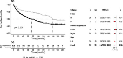
3 RESULTSThe mean age of the four patients with a history of pulmonary surgery was 58.50 ± 2.38 years, and the male to female ratio was 1:1. Lobectomy was performed in three patients, and wedge resection and segmentectomy in one patient; all patients received thoracoscopic surgery. Three patients were diagnosed with non-small cell lung cancer, one with sclerosing pulmonary cell tumor. None of the patients received radiotherapy or chemotherapy after surgery. All patients presented fever, and all patients had body temperature > 38°C. The onset time of COVID-19 was from 3 to 10 days after surgery, with a median time of 6.75 days. Other symptoms were accompanied with fever, including nonproductive cough (two), rigors (one), chest distress (one), chest pain (one), productive cough (one), myalgia (one), and diarrhea (one). One patient died and three recovered and discharged from the hospital. The basic information and clinical characteristics of the four patients are shown in Table 1.
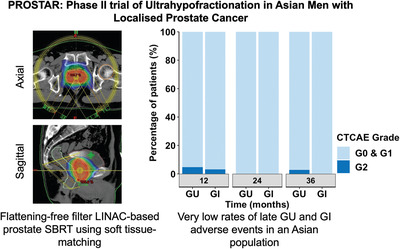
TABLE 1. Clinical characteristics, treatment, and outcomes of patients with lung surgery Patient 1 Patient 2 Patient 3 Patient 4 Gender Male Female Male Female Age 61 60 57 56 Operation LUL lobectomy LUL wedge resection +LLL segmentectomy RUL lobectomy RUL lobectomy Histological type SCC Ade + Ade Ade PSP Pathological stage pT2N2M0, IIIA pT1bN0M0, IA pT1bN0M0, IA NA Radiotherapy No No No NA Chemotherapy No No No NA Surgery to onset of symptoms 3 days 7 days 7 days 10 days Highest temperatures, ℃ 38.2 38.3 38.5 38.4 Headache No No No No Chills No No No No Rigors No Yes No No Cough Yes No No Yes Productive cough Yes No No No Sore throat No No No No Chest distress Yes No Yes No Chest pain No No Yes No Dyspnea No No Yes No Fatigue No No No No Myalgia Yes No No No Diarrhea No No No Yes Antiviral therapy Yes Yes Yes Yes Antibiotics Yes Yes Yes Yes TCM Yes Yes Yes Yes Immunoglobulin Yes No Yes Yes Systemic corticosteroids Yes No Yes Yes Oxygen therapy High-flow oxygen No No No Yes NMV No No No Yes IMV No No No Yes Outcome Discharge Discharge Discharge Death Abbreviations: Ade, adenocarcinoma; LLL, left lower lobe; LUL, left upper lobe; NA, not applicable; PSP, pulmonary sclerosing pneumocytoma; RLL, right lower lobe; RUL, right upper lobe; SCC, squamous cell carcinoma; TCM, traditional Chinese medicine.The chest computer tomography (CT) manifestations of the four patients are shown in Figure 2, all patients showed ground-glass opacity or patchy shadowing and the infection was predominant in the lobe contralateral to the side of surgery in three patients. The symptoms gradually improved and patchy shadow has been absorbed in three patients. In patient 4, lung effusions aggravated as disease deteriorated; despite receiving mechanical ventilation, the patients eventually died from respiratory failure on the 42th postoperative day.
 CT manifestations in four Patients with COVID-19 after lung surgery. (A) A 61-year-old male underwent VATS left upper lobectomy and lymphadenectomy. CT images on day 10 postoperation show patchy shadow in the right lung. CT images on day 23 postoperation show the shadows have been absorbed. (B) A 60-year-old female underwent VATS wedge resection of left upper lobe with basal segmentectomy and lymphadenectomy. CT images on day 16 postoperation show patchy shadow in right lower lobe. CT images on day 36 postoperation show the patchy shadow has been absorbed.(C) A 57-year-old male underwent VATS right upper lobe lobectomy and lymphadenectomy. CT images on day 9 postoperation show patchy shadow in left lower lobe. CT images on day 45 postoperation show the patchy shadow has been absorbed.(D) A 56-year-old female underwent VATS right lower lobe lobectomy. CT images on day 16 postoperation show bilateral multifocal ground-glass opacities, and x-ray on day 33 postoperation show aggravated exudation of the left lung and right pneumothorax [Colour figure can be viewed at wileyonlinelibrary.com]
The comparison of demographic information between the two groups is shown in Table 2. Incidence of hypertension in the surgical and nonsurgical groups (1 of 4 [25.0%] vs 5 of 16 [31.3%]; P = 1.0) showed no statistical difference, and the participants of neither group had diabetes, coronary heart disease, or cerebrovascular disease. The mortality rate of the surgical group (1/4, 25.0%) was higher than that of the nonsurgical group (1/16, 6.3%).
TABLE 2. Demographic of the two groups Variables Lung surgery group (n = 4) Nonsurgery group (n = 16) P Number of patients, n (%) 4 (20) 16 (80) Gender (male/female) 2/2 8/8 1.0 Age, mean (SD), years 58.50 (2.38) 58.56 (3.20) .97 Pre-existing comorbidities Hypertension, n (%) 1 (33.3) 5 (29.2) 1.0 Diabetes mellitus, n (%) 0 0 Coronary artery disease, n (%) 0 0 Cerebrovascular disease, n (%) 0 0We compared the laboratory indexes of the two groups (Figure 3). Due to the limitation of sample size, we did not conduct statistical analysis. However, we still find that albumin, hemoglobin, lymphocytes, IL-6, and ferritin were lower in the surgery group.
 The laboratory indexes of patients with COVID-19 in nonsurgery group and lung surgery group. The values of albumin, ferritin, IL-6, hemoglobin, lymphocytes of patients with COVID-19 in nonsurgery group (red), and lung surgery group (blue) on admission are presented [Colour figure can be viewed at wileyonlinelibrary.com]
4 DISCUSSION
The laboratory indexes of patients with COVID-19 in nonsurgery group and lung surgery group. The values of albumin, ferritin, IL-6, hemoglobin, lymphocytes of patients with COVID-19 in nonsurgery group (red), and lung surgery group (blue) on admission are presented [Colour figure can be viewed at wileyonlinelibrary.com]
4 DISCUSSION
Peng et al10 found that the death rate of 11 patients with COVID-19 after chest surgery was 27.3%; Cai et al11 reported a mortality rate of 28.6% in seven patients undergoing lung surgery. In our study, the mortality rate was 25.0% in the surgical group, which was much higher than the 6.3% in the nonsurgical group. These results may be associated with the loss of lung function and decreased immune function following lung surgery.
SARS-CoV-2 uses angiotensin-converting enzyme II (ACE-2) as a cell entry receptor.12 ACE-2 is expressed in a variety of different tissues, including upper and lower respiratory tract, myocardium, and gastrointestinal mucosa.13 Because of the high expression of ACE-2 in the lung tissue, the lungs are mainly affected in COVID-19. The pathological findings of patients with COVID-19 showed a decrease in lung cells and the formation of a clear membrane, suggesting ARDS.14, 15 Since adult lung stroma is not renewable, lung function will inevitably be lost after lung surgery. Gu et al5 found that forced expiratory volume in 1 second decreased by 13.58% (±9.98) 3 months after lobectomy, and by 8.49% (±10.36) 1 year after lobectomy. The carbon monoxide diffusing capacity decreased by 14.56% (±13.35) 3 months after lobectomy, and by 8.86% (±14.03) 1 year after lobectomy. Kim et al6 and Nomori et al7 reported similar results. The loss of lung function theoretically depends on the amount of lung tissue removed during surgery.16 Peng et al10 reported that the number of pulmonary segmentations greater than or equal to five was found to be associated with death by COVID-19. In this study, all patients had more than three lung segments removed, and developed COVID-19 symptoms within 10 days after surgery. Although we did not confirm the lung function level of the patients by pulmonary function examination, it was predicted that the lung function level of the surgical group was lower than that of the normal population, which could affect the prognosis.
The decrease in immune function after surgery can be evaluated by the decrease in lymphocytes and increase in inflammatory factors. Lymphocytosis often occurs immediately after lung surgery and is associated with postoperative pneumonia.17 Ogawa et al. found that it takes 2 weeks for peripheral blood lymphocyte function to recover after surgery.18 In this study, all patients developed COVID-19 within 2 weeks after surgery. Although the absolute value of lymphocytes of the surgical group was lower than the nonsurgical group, lymphocytopenia is a characteristic of SARS-CoV-2 infection and indicates the severity of the disease.3, 19 Moreover, postoperative lymphocytic decline could complicate the conditions.
Cytokine storms may be the pathogenesis of COVID-19, and the elevation of IL-6 indicates the level of inflammatory response.20 Whitson et al21 found that the level of IL-6 significantly increased after thoracotomy and reached a peak on the first day after surgery, but did not increase significantly in the thoracoscopic surgery group. Wolf et al22 found similar results. In this study, there was not a higher IL-6 levels in the surgical groups. Similarly, other indicators reflecting the severity of COVID-19, such as ferritin,23 also did not get a higher level.
Majority of patients had undergone lung surgery for lung cancer. In our study, three (75%) patients had lung cancer. Zhang et al24 showed that 35.7% of 28 cancer patients with COVID-19 developed severe life-threatening complications, and 28.6% died. The authors suggest that immunosuppression in tumor patients is associated with a poorer prognosis. Mehta et al25 also reported similar results.
5 CONCLUSIONPatients contracting COVID-19 after lung surgery presented a higher death rate; hence, it is necessary to omit lung surgery in patients with active COVID-19 infection.
6 LIMITATIONSThis was a single-center retrospective study with a small sample size, and the results need to be verified by a large sample size study. Heterogeneity existed in four patients undergoing lung surgery, not all of which were tumor patients. Most of the patients were hospitalized after a considerably long duration, and most had taken antiviral and antibacterial drugs outside the hospital, which might have influenced the signs and test results. The proportion of critically ill patients in our wards was high, which might have influenced the results of the analysis.
ACKNOWLEDGMENTSThe authors thank all the healthcare workers from the isolation critical care wards in Tongji Hospital, Tongji Medical College, Huazhong University of Science and Technology. Project of Development Center for Medical Science and Technology, National Health Commission of the PRC; Grant Number: W2017ZWS17.
AUTHOR CONTRIBUTIONSQinggang Ge and Shaohua Ma participated in the design of the study. Jie Bai contributed to the collection, analysis, and interpretation of data. Hongling Chu performed the statistical analysis.
CONFLICT OF INTERESTAll authors declare no conflict of interest.
ETHICAL STATEMENTThis study was approved by the ethics committee of Peking University Third Hospital (IRB00006761-M2020060).
After publication, we can provide the data to others with the permission of the corresponding author. A proposal with detailed description of study objectives and statistical analysis plan will be needed for evaluation of the reasonability of requests. The corresponding authors have the right to decide whether to share the data or not based on the research objectives and plan provided.
REFERENCES
1 WHO. WHO Director-General's opening remarks at the media briefing on COVID-19. March 2020. https://www.who.int/dg/speeches/detail/who-director-general-s-opening-remarks-at-themedia-briefing-on-covid-19—11-march-2020. Accessed March 30, 2020. 2Huang C, Wang Y, Li X, et al. Clinical features of patients infected with 2019 novel coronavirus in Wuhan, China. Lancet. 2020; 395: 497- 506. 3Chen N, Zhou M, Dong X, et al. Epidemiological and clinical characteristics of 99 cases of 2019 novel coronavirus pneumonia in Wuhan, China: a descriptive study. Lancet. 2020; 395: 507- 513. 4Wang D, Hu B, Hu C, et al. Clinical characteristics of 138 hospitalized patients with 2019 novel coronavirus-infected pneumonia in Wuhan, China. JAMA. 2020. https://doi.org/10.1001/jama.2020.1585. 5Gu Z, Wang H, Mao T, et al. Pulmonary function changes after different extent of pulmonary resection under video-assisted thoracic surgery. J Thorac Dis. 2018; 10: 2331- 2337. 6Kim SJ, Lee YJ, Park JS, et al. Changes in pulmonary function in lung cancer patients after video-assisted thoracic surgery. Ann Thorac Surg. 2015; 99: 210- 217. 7Nomori H, Shiraishi A, Cong Y, Sugimura H, Mishima S. Differences in postoperative changes in pulmonary functions following segmentectomy compared with lobectomy. Eur J Cardiothorac Surg. 2018; 53: 640- 647. 8Liang W, Guan W, Chen R. Cancer patients in SARS-CoV-2 infection: a nationwide analysis in China. Lancet Oncol. 2020. https://doi.org/10.1016/S1470-2045(20)30096-6. 9Li YK, Peng S, Li LQ, et al. Clinical and transmission characteristics of Covid-19: a retrospective study of 25 cases from a single thoracic surgery department. Curr Med Sci. 2020; 40: 295- 300. 10Peng S, Huang L, Zhao B, et al. Clinical course of coronavirus disease 2019 in 11 patients after thoracic surgery and challenges in diagnosis. J Thorac Cardiovasc Surg. 2020. https://doi.org/10.1016/j.jtcvs.2020.04.005. 11Cai Y, Hao Z, Gao Y, et al. Coronavirus disease 2019 in the perioperative period of lung resection: a brief report from a single thoracic surgery department in Wuhan, People's Republic of China. J Thorac Oncol. 2020. https://doi.org/10.1016/j.jtho.2020.04.003. 12Zhou P, Yang XL, Wang XG, et al. A pneumonia outbreak associated with a new coronavirus of probable bat origin. Nature. 2020; 579: 270- 273. 13Harmer D, Gilbert M, Borman R, Clark KL. Quantitative mRNA expression profiling of ACE 2: a novel homologue of angiotensin converting enzyme. Febs Lett. 2002; 532: 107- 110. 14Tian S, Hu W, Niu L, Liu H, Xu H, Xiao SY. Pulmonary pathology of early-phase 2019 novel coronavirus (COVID-19) pneumonia in two patients with lung cancer. J Thorac Oncol. 2020. https://doi.org/10.1016/j.jtho.2020.02.010. 15Xu Z, Shi L, Wang Y, et al. Pathological findings of COVID-19 associated with acute respiratory distress syndrome. Lancet Respir Med. 2020; 8: 420- 422. 16Ueda K, Hayashi M, Tanaka N, Tanaka T, Hamano K. Long-term pulmonary function after major lung resection. Gen Thorac Cardiovasc Surg. 2014; 62: 24- 30. 17Dupont G, Flory L, Morel J, et al. Postoperative lymphopenia: an independent risk factor for postoperative pneumonia after lung cancer surgery, results of a case-control study. Plos One. 2018; 13:e205237. 18Ogawa K, Hirai M, Katsube T, et al. Suppression of cellular immunity by surgical stress. Surgery. 2000; 127: 329- 336. 19Yang X, Yu Y, Xu J, et al. Clinical course and outcomes of critically ill patients with SARS-CoV-2 pneumonia in Wuhan, China: a single-centered, retrospective, observational study. Lancet Respir Med. 2020. https://doi.org/10.1016/S2213-2600(20)30079-5. 20Zhang C, Wu Z, Li JW, Zhao H, Wang GQ. The cytokine release syndrome (CRS) of severe COVID-19 and interleukin-6 receptor (IL-6R) antagonist Tocilizumab may be the key to reduce the mortality. Int J Antimicrob Agents. 2020. https://doi.org/10.1016/j.ijantimicag.2020.105954. 21Whitson BA, D'Cunha J, Andrade RS, et al. Thoracoscopic versus thoracotomy approaches to lobectomy: differential impairment of cellular immunity. Ann Thorac Surg. 2008; 86(6): 1735- 1744. 22Wolf J, Rose-John S, Garbers C. Interleukin-6 and its receptors: a highly regulated and dynamic system. Cytokine. 2014; 70: 11- 20. 23Chen G, Wu D, Guo W, et al. Clinical and immunologic features in severe and moderate coronavirus disease 2019. J Clin Invest. 2020. https://doi.org/10.1172/JCI137244. 24Zhang L, Zhu F, Xie L, et al. Clinical characteristics of COVID-19-infected cancer patients: a retrospective case study in three hospitals within Wuhan, China. Ann Oncol. 2020. https://doi.org/10.1016/j.annonc.2020.03.296. 25Mehta V, Goel S, Kabarriti R, et al. Case fatality rate of cancer patients with COVID-19 in a New York hospital system. Cancer Discov. 2020. https://doi.org/10.1158/2159-8290.CD-20-0516.
留言 (0)