Table S1. NMR data for atramycin C (1) in DMSO-d6a
Table S2. NMR data foremycin G (2) in DMSO-d6a
Figure S1. 1H NMR spectrum (600 MHz) of atramycin C (1) in DMSO-d6.
Figure S2. 13C NMR spectrum (150 MHz) of atramycin C (1) in DMSO-d6.
Figure S3. DEPT-135 spectrum (150 MHz) of atramycin C (1) in DMSO-d6.
Figure S4. COSY spectrum (600 MHz) of atramycin C (1) in DMSO-d6.
Figure S5. HSQC spectrum (600 MHz) of atramycin C (1) in DMSO-d6.
Figure S6. HMBC spectrum (600 MHz) of atramycin C (1)in DMSO-d6.
Figure S7. NOESY spectrum (600 MHz) of atramycin C (1) in DMSO-d6.
Figure S8. HRESIMS spectrum of atramycin C (1).
Figure S9. 1H NMR spectrum (600 MHz) of emycin G (2) in DMSO-d6.
Figure S10. 13C NMR spectrum (150 MHz) of emycin G (2) in DMSO-d6.
Figure S11. DEPT-135 spectrum (150 MHz) of emycin G (2) in DMSO-d6.
Figure S12. COSY spectrum (600 MHz) of emycin G (2) in DMSO-d6.
Figure S13. HSQC spectrum (600 MHz) of emycin G (2) in DMSO-d6.
Figure S14. HMBC spectrum (600 MHz) of emycin G (2) in DMSO-d6.
Figure S15. NOESY spectrum (600 MHz) of emycin G (2) in DMSO-d6.
Figure S16. HRESIMS spectrum of emycin G (2).
Figure S17. HPLC spectra of the a) derivatized standard L -rhamnose,b) derivatized standard D -rhamnose, and c) derivatized product of hydrolysed sugar from atramycin C (1), respectively.
Figure S18. LC/MS analysis for the a) derivatized standard L-rhamnose, b) derivatized standard D-rhamnose.
Figure S19. LC/MS analysis for the derivatized product of hydrolysed sugar from atramycin C (1).
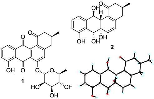
Table S3. Crystal data and structure refinement for compound 2
Table S4. Crystal data and structure refinement for compound 3
Table S5. Crystal data and structure refinement for compound 4


留言 (0)