
記住我


Medicinal plants have been of great importance in the management of human diseases. The finding that even our closest living relatives, the chimpanzees, have developed a culture to use specific plants for healing purposes (Page et al., 1992), indicates that, from prehistoric time, humankind have used plants for healing. Classical examples are the Neem tree (Azadirachta indica) (reviewed in Kumar and Navaratnam, 2013), and the bark of the willow tree (Salicis cortex) (for reviews see Jack, 1997; Mahdi, 2010). To date, half a million of plant secondary metabolites with known biological activities are considered as a sustainable source of novel drug candidates for the pharmacological treatment of various diseases (recent reviews in Demain and Vaishnav, 2011; David et al., 2015; Shen, 2015; Bernardini et al., 2018). Numerous effective drugs with economic impact are of herbal origin, such as the alkaloids morphine (Papaverum somniferum L.), paclitaxel (Taxus brevifolia), digitoxin (Digitalis purpurea), curare (Chondrodendron tomentosum), or the sesquiterpene artemisinin (qinghaosu 青蒿素 in Chinese, Artemisia annua), are still isolated from herbal plants (for reviews see Mahdi, 2010; Shen, 2015; Bernardini et al., 2018). Others, such as aspirin (acetyl-salicylic acid), while being meanwhile synthetized technically, derive from plants as well (Salix sp.). Thus, the impact of pharmacology of natural products from medicinal plants for the discovery of novel drugs is considerable, reviving the interest in traditional systems of healing, but also fueling controversies about the intellectual property linked with this traditional knowledge (Fredriksson, 2017).
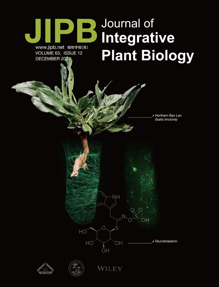
Traditional Chinese medicine (TCM) belongs to the most elaborate systems of healing (Waller, 1998). The compendium Bencao Gangmu by Shizhen Li, published in 1578, lists more than 10,000 recipes, based on 1,892 plant species, forms the base of the current TCM pharmacopeia (Xu et al., 2019). This extensive body of traditional knowledge represents a huge, still almost untapped, reservoir for the discovery of new bioactive compounds. In the context of the current Covid-19 pandemic, TCM plants with antiviral activity have attracted attention. Northern Ban Lan is traditionally used for its antiviral activity, but also to cure a broad spectrum of other diseases (for review see Hamburger, 2002) due to its anti-inflammatory and anti-microbial effect (Recio et al., 2006a, 2006b; Brattström et al., 2010; Ullah et al., 2017). While true Northern Ban Lan is supposed to be Isatis tinctoria (Dyer's woad), often the more easily accessible, closely related I. indigotica (indigo) is used as a substitute. Both the root (I. radix, Ban Lan Gen, 板蓝根 in Chinese), and the leaf (I. folia, Da Qing Ye, 大青叶 in Chinese) are used for the clinical treatment of virus-related respiratory diseases such as severe acute respiratory syndrome (SARS), and H1N1 (Lin et al., 2005; Wang et al., 2011). Currently, Northern Ban Lan is subject to several clinical trials for therapies against Covid-19 (for a recent review see Yang et al., 2020).
The medical activities of Isatis extract are thought to depend on the synergistic effect of multiple components (Zhou and Zhang, 2013). Over the last decades, more than 100 bioactive constituents have been identified in methanolic extracts of I. tinctoria leaves and roots, including numerous alkaloids, fatty acids, and carotenoids (Mohn et al., 2009), as well as phenylpropanoid derivatives, such as flavonoids, hydroxyl-cinnamic and hydroxyl-benzoic acid derivatives, or mono- and oligo-lignols (Nguyen et al., 2017). Especially, the glucosinolates, indole-derived compounds, have attracted interest for their prospective medical properties (Mohn and Hamburger, 2008; Nguyen et al., 2017). For instance, indirubin, a bis-indole alkaloid, was reported to mitigate expression of the influenza infection marker RANTES (regulated on activation, normal T cell expressed and secreted) in a bronchitis cell model (Mak et al., 2004; Sethi et al., 2006). Although these phenomena appear as quite distant, they are functionally linked, namely by microtubules: influenza viruses hijack the microtubules of their hosts for intracellular movement, uncoating, and exit from the cell (for a recent review see Simpson and Yamauchi, 2020). Also the transcription factor nuclear factor-κB is tethered to microtubules and can, upon modulation of microtubule dynamics, activate apoptosis (Rai et al., 2015).
Microtubules are dynamic components of the cytoskeleton in eukaryotic cells, which are essential for various cellular functions, such as development and maintenance of cell shape, intracellular transport of vesicles and other components, cell signaling and cell division (see reviews in Dumontet and Jordan, 2010; Forth and Kapoor, 2017; Lu and Gelfand, 2017; Muroyama and Lechler, 2017; Dogterom and Koenderink, 2019). Interference with microtubular dynamics by small-molecule inhibitors leads to mitotic arrest and can trigger apoptosis (reviewed in Peterson and Mitchison, 2002; Florian and Mitchison, 2016). For this reason, anti-microtubular compounds are among the most effective drugs used in the treatment of solid tumors and hematological malignancies (for reviews see Jordan et al., 1998; Kaul et al., 2019). Over the past decades, a large number of microtubule-targeting compounds have been discovered during large-scale screens of natural products, and obtained approval for cancer therapy (reviewed in Altmann and Gertsch, 2007; Florian and Mitchison, 2016; Mahaddalkar and Lopus, 2017). Furthermore, microtubules are used for the transport of RNA, which is a central element of morphogenesis. A classic example is the microtubule-dependent transport of maternal messenger RNA (mRNA) during polarization of the Drosophila embryo (St Johnston and Nüsslein-Volhard, 1992), or the long-distance transport of mRNA in neurons (reviewed in Glock et al., 2017). Several RNA viruses hijack this essential function for their own purpose, for instance to move from the plasma membrane to the nuclear envelope (Wang et al., 2017). Anti-microtubule agents might, therefore, affect virus infection. The fact that also plant microtubules are usurped for transport of RNA viruses (tobacco mosaic virus: Heinlein et al., 1995; grapevine fan leaf virus: Laporte et al., 2003), indicates that, here, an evolutionary ancient target is exploited, which is shared between plants and animals.
However, not only viruses, but also plants themselves, use microtubules as targets for chemical warfare. For instance, the monoterpene citral is used to suppress germination of competing plants by blocking the division spindle (Chaimovitsh et al., 2010), and several alkaloids serve to ward off herbivores, by affecting integrity or dynamics of their microtubules. Among those, Vinca alkaloids and taxanes are of medical relevance, because they are widely used for tumor therapy (Nogales et al., 1995; Jordan et al., 1998). While being relatively conserved, tubulins of plants and animals also have evolved minor, but specific differences that lead to specificity of such plant-derived anti-microtubule compounds (for review see Dostál and Libusová, 2014). Nevertheless, in contrast to the microtubule-associated proteins, the molecular conservation of tubulins is very high – for instance, porcine brain tubulin can be microinjected into living plant cells and integrates readily with the host cytoskeleton and then participates normally even in very specific responses (Himmelspach et al., 1999).
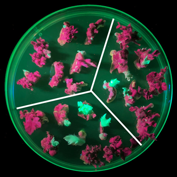
Based on the conservation of tubulins between animals and plants, we ventured to search for anti-microtubule compounds from I. tinctoria (Northern Ban Lan) using activity-guided high-performance liquid chromatography (HPLC) fractionation as strategy. We deliberately chose a plant-cell system as readout for bioactivity. The use of a tobacco cell line, where microtubules are labeled by green fluorescent protein (GFP)-tagged plant tubulin (Kumagai et al., 2001), not only allowed us to follow the response of microtubules in vivo, but also selected for activities that are directed to targets shared between mammalian and plant cells: While the microtubule-associated proteins are quite divergent between these two cell types, a bioactivity seen on plant microtubules is most likely acting on the level of the evolutionarily conserved tubulins themselves. Using high-resolution mass spectrometry (HRMS) and tandem mass spectrometry (MS/MS), our search identified glucobrassicin as an anti-microtubule compound from the TCM plant Northern Ban Lan (I. tinctoria).
RESULTS Anti-microtubular activity of I. tinctoria roots originates from a specific fractionLeaf and root material from I. tinctoria was extracted with two different solvent systems, either MeOH/H2O 8:2, or ethyl acetate (EtOAc) (yields are shown in Table S1, representative liquid chromatography ultra violet chromatograms in Figure S1). These crude extracts were fractionated at fixed retention time (tR) intervals, and the resulting fractions were screened for their ability to induce a response of GFP-tagged microtubules in tobacco Bright Yellow 2 (BY-2) cells expressing GFP-NtTuA3 (Nicotiana tabacum α-tubulin-3) as readout for bioactivity. According to the differentiation of extraction material (leaf, root) and solvent (MeOH, EtOAc), the total 54 fractions were named ML-1 to ML-14 (MeOH/H2O 8:2 leaf fractions), EL-1 to EL-12 (EtOAc leaf fractions), MR-1 to MR-14 (MeOH/H2O 8:2 root fractions), and ER-1 to ER-14 (EtOAc root fractions), respectively (Table S3).
These fractions were then administered to expanding cells of microtubular marker line TuA3-GFP to probe for potential effects upon cortical microtubules. In the solvent control, microtubules were arranged in parallel bundles aligned perpendicular to the expansion axis of the cell, as it is typical for elongating cells (Figure S2). In fact, some fractions induced perturbations in the integrity and the organization of cortical microtubules, while others did not produce any effect. For instance, out of the 14 fractions obtained by methanolic extraction of leaves, only three (ML-4, ML-5, and ML-10) were able to induce microtubular perturbations (Figure S2, white arrows). This effect was relatively swift, as shown by a time course experiment (Figure S3). For the more active fractions ML-5 and ML-10, a clear disassembly of cortical microtubules was visible already from 15 min after application. Considering the EtOAc leaf extracts, two fractions (EL-3, EL-9) produced the microtubule elimination (Figure S4). Also these fractions affected microtubules strongly from 15 min after application (Figure S5). Methanolic extraction of the roots yielded one fraction (MR-1), which was more active with respect to microtubule elimination than any other fraction recovered in this study (Figure S6). As early as 10 min, most of the microtubules had disappeared (Figure S7). However, extraction of roots with EtOAc yielded only one fraction (ER-9) with a very mild effect (Figure S8). This mild effect became evident from around 20 min (Figure S9). The microtubule activities of the different fractions are summarized in Table S5: In total, three MeOH/H2O 8:2 (ML-4, ML-5, and ML-10), two EtOAc fractions (EL-3, EL-9) from leaves were screened out. For roots only one MeOH/H2O 8:2 (MR-1) and one EtOAc fraction (ER-9) displayed anti-microtubular activity. Among these seven active fractions, MR-1 induced the strongest microtubule response and was therefore put into the focus of subsequent analysis.
The most active fraction MR-1 was as efficient as the anti-microtubular herbicide Oryzalin (Figure 1A). A quantification of microtubule integrity revealed that none of the other root fractions caused any significant response of microtubules (Figure 1B). This response was also seen for dilutions of MR-1 (Figure 2). For instance, a dilution of 1:5 made microtubules disappear from as early as 3 min. As early as 10 min, most microtubules had been eliminated, and only punctate remnants of microtubules were seen, probably corresponding to microtubule organizing centers (MTOCs). For the 1–10 dilution, there was no observable change over the first 15 min, but from 20 min some fluorescent dots appeared, indicative of a slight effect upon microtubules.

Response of microtubules to treatment with fractions obtained from MeOH/H2O 8:2 extracts of Isatis tinctoria roots in the microtubule marker line Nicotiana tabacum L. cv Bright Yellow 2 α-tubulin-3 green fluorescent protein (BY-2 TuA3-GFP)
(A) Representative cells after 60 min of treatment with the solvent control (1% dimethyl sulfoxide), the anti-microtubular herbicide Oryzalin as positive control, and fraction MR-1 as compared to untreated cells observed by spinning-disc microscopy. Geometrical projections from z-stacks collected at intervals of 0.5 µm are shown. (B) Quantification of microtubule integrity for the 14 methanolic fractions obtained from I. tinctoria root (MR-1 to MR-14), as compared to untreated cells, control, solvent control, and the positive control Oryzalin. ***Difference significant at P < 0.001 based on a t-test.

Time-lapse study of the microtubular response to a 1:5 dilution (upper row) and a 1:10 dilution (lower row) of the methanolic root fraction MR-1 in expanding cells of the microtubule marker line Nicotiana tabacum L. cv Bright Yellow 2 α-tubulin-3 green fluorescent protein (BY-2 TuA3-GFP) followed over 30 min
Geometrical projections from z-stacks collected at intervals of 0.5 µm are shown.
The effect of fraction MR-1 is specific for microtubulesThe efficient elimination of microtubules by the most active fraction MR-1 might be an unspecific consequence of a potential cytotoxicity. We checked, therefore, whether MR-1 would also disrupt actin filaments, as a second element of the plant cytoskeleton, and whether this fraction exerts phytotoxic effects. For this purpose, we employed the actin marker line GF11 (Sano et al., 2005), where actin filaments are tagged by the actin-binding domain 2 of plant fimbrin in fusion with GFP. Even after treatment with MR-1 for 60 min in the same dilutions as for the microtubule experiment (Figure 2), the actin cytoskeleton remained fully developed (Figure 3), no matter whether the more concentrated 1:5 dilution (Figure 3B), or the 1:10 dilution (Figure 3C) were used. These observations were consistent with the quantification results (Figure S10). However, there was an effect of the treatment: as compared to the solvent control (Figure 3A), actin filaments appeared more bundled. However, they retained their full integrity, which contrasts with the thorough elimination observed for microtubules observed for the same treatments, and even for much earlier time points (Figure 2). Thus, MR-1 does not cause a generic breakdown of the cytoskeleton, but is specific for microtubules.

Response of actin filaments to the methanolic root fraction MR-1 in expanding cells of the actin marker line Nicotiana tabacum L. cv Bright Yellow 2 α-tubulin-3 green fluorescent protein (BY-2 TuA3-GFP) 30 min after application
(A) Solvent control, (B) 1:5 dilution, (C) 1:10 dilution. Geometrical projections from z-stacks collected at intervals of 0.5 µm are shown for representative cells.
Since fraction MR-1 targeted to microtubules and, therefore, might induce mitotic arrest followed by programmed cell death, potential mortality induced by two dilutions from fraction of MR-1 was scored for 6, 12 and 24 h using the Evans Blue dye exclusion test in tobacco BY-2 cells. Since only a limited amount of the fraction was available, and since mortality scoring requires a larger suspension volume, the dilutions had to be increased (1:200 and 1:1,000, respectively) as compared to the microscope tests. Even so, the more concentrated dilution of 1:200 dilution required 24 h to induce a moderate mortality of around 30% (Figure 4). Thus, there seems to be no acute cytotoxicity, but the slow progression of mortality over 1 d is consistent with a working hypothesis, where mortality is caused by mitotic arrest, since the doubling time of BY-2 cells is in the range of 1 d (Rajabi et al., 2017).

Time course of cell mortality in response to dilutions of fraction MR-1
The relative frequency of dead cells after treatment with MR-1 as compared to control (white bars) in Nicotiana tabacum L. cv Bright Yellow 2 (BY-2) was followed over time scoring samples of 1 500 cells for each data point. Data represent three independent biological replicates.
UHPLC-HRMS analysis for identification of main constituents in fraction MR-1MR-1, the most polar fraction obtained by extraction of I. tinctoria roots with MeOH/H2O 8:2, showed the strongest activity against microtubules (Figures 1, 2). Due to the complexity of the sample and the low amounts available, the MR-1 fraction was investigated by UHPLC-(–) electrospray ionization (ESI)-HRMS and MS/MS (Figure 5). The resulting data revealed the presence of 16 compounds (Table S4). Since epigoitrin ((R)–goitrin, (R)-5-vinyloxazolidine-2-thione) and goitrin ((S)–goitrin, (S)-5-vinyloxazolidine-2-thione) have been described as markers of antiviral efficacy in I. radix (Ban Lan Gen) in the Chinese Pharmacopoeia 2015 (Dan et al., 2016), their commercially available glucosinolate precursors epiprogoitrin (allylic glucosinolate), progoitrin (allylic glucosinolate), and, glucobrassicin (indolic glucosinolate) were selected and considered in more detail as potential candidates for the anti-microtubule activity. Also, indirubin, a potential anticancer component of I. tinctoria, proposed to bind to tubulin or inhibit several kinases involved in cell division and exerting antimitotic functions against HeLa cells (Mohan et al., 2018), was included into further analysis.

Ultra-high-performance liquid chromatography electrospray ionization high-resolution mass spectrometry (HPLC-(–) ESI-HRMS) of fraction MR-1 obtained from I. tinctoria root extraction with MeOH/H2O 8:2
The total ion chromatogram (TIC) is shown on the top, the extracted ion chromatogram (EIC of theoretical [M–H]− ions) of epiprogoitrin (C11H19NO10S2), progoitrin (C11H19NO10S2), and glucobrassicin (C16H20N2O9S2) on the bottom.
Glucobrassicin exerts anti-microtubule activityTo find out which component of fraction MR-1 is responsible for the anti-microtubule effect, we assessed the response of BY-2 TuA3 cells to standard compounds of epiprogoitrin, progoitrin, glucobrassicin, and indirubin in a concentration of 50 µmol/L after 60 min. Upon visualization of microtubules using spinning-disc microscopy (Figure 6), glucobrassicin clearly stuck out, because it strongly eliminated microtubules accompanied by an increase of diffuse fluorescence in the cytoplasm (Figure 6E). In contrast, neither epiprogoitrin, nor progoitrin produced any effect exceeding that seen for the solvent control (compare Figure 6C, D to Figure 6B). Likewise, there was no obvious difference between solvent control and indirubin treatment (compare Figure 6E to Figure 6B). The quantification of microtubule integrity indicated that only glucobrassicin induced significant microtubule responses (Figure S11). We tested, whether a combination of 50 µmol/L epiprogoitrin and progoitrin was able to eliminate microtubules, but observed only a mild effect, manifest as increased diffuse background fluorescence if compared to the solvent control (Figure S12A, B). If we raised the concentration to 500 µmol/L for epiprogoitrin, progoitrin, or indirubin, we evoked an obvious elimination of microtubules for epiprogoitrin, but not for progoitrin (Figure S12C, B). Likewise, 500 µmol/L of indirubin were not effective in eliminating microtubules (Figure S12D). Further, an exemplary time course of microtubule responses for glucobrassicin was observed and checked in the treatments of 30, 60, 120 and 180 min treatment. The results showed that the integrity of microtubules continued to decline with time (compare Figure 7E to Figure 7B). After 120 min, the microtubule network almost disappeared (Figure 7D, F). Thus, glucobrassicin exerts a specific and clear anti-microtubular activity, which is not seen in any of the other compounds tested.

Appearance of microtubules in the microtubule marker line Nicotiana tabacum L. cv Bright Yellow 2 α-tubulin-3 green fluorescent protein (BY-2 TuA3-GFP) prior to treatment (A), in response to the solvent (1% dimethyl sulfoxide; B), epiprogoitrin (50 µmol/L; C), progoitrin (50 µmol/L; D), glucobrassicin (50 µmol/L; E), and indirubin (50 µmol/L; F) after 60 min of treatment observed by spinning-disc microscopy
Geometrical projections from z-stacks collected intervals of 0.5 µm are shown. Lower row shows zoom-ins of the region marked by the white box in the upper row.

Exemplary time course of microtubule responses for glucobrassicin in the microtubule marker line Nicotiana tabacum L. cv Bright Yellow 2 α-tubulin-3 green fluorescent protein (BY-2 TuA3-GFP)
The cellular microtubule prior to treatment (A), and in response to 50 µmol/L glucobrassicin treatment in a time course of 30 min (B), 60 min (C), 120 min (D), and 180 min (E) were observed by spinning-disc microscopy. Geometrical projections from z-stacks collected at intervals of 0.5 µm are shown. Lower row shows zoom-ins of the region marked by the white box in the upper row. (F) Quantification of microtubule integrity for the glucobrassicin in different time points.
If glucobrassicin would act on microtubule-associated proteins that differ fundamentally between plant and animal cells, it should not affect microtubules in mammalian cells. We followed, therefore, the response of HeLa cells expressing a GFP-tubulin marker to 50 µmol/L of glucobrassicin over 90 min (Figure 8), but also assessed the cellular response after 1 d (Figure 9). Microtubules were strongly affected already at 30 min after addition of glucobrassicin (Figure 8C) and even after 90 min had still not returned to the situation seen in the solvent control (Figure 8A, B). In agreement with our observations, the quantification results also revealed that glucobrassicin significantly reduced microtubule integrity (Figure S13). In addition, after treatment with 25 µmol/L of glucobrassicin after 24 h, many cells were found to have entered apoptosis, which was even more advanced as that induced by 1 µmol/L colchicine, used as a positive control (Figure 9). Thus, the anti-microtubular effect of glucobrassicin was confirmed in HeLa cells as well, and this was followed by a drastic apoptosis in these mammalian cells.

Exemplary time course of microtubule responses for glucobrassicin in HeLa cells expressing a green fluorescent protein (GFP)-tubulin marker
The cellular microtubule of control treatment 0 min (A) and 60 min (B), and in response to 50 µmol/L glucobrassicin in an exemplary time course of 30 min (C), 90 min (D) were observed by spinning-disc microscopy. Geometrical projections from z-stacks collected at intervals of 0.5 µm are shown. Lower row shows zoom-ins of the region marked by the white box in the upper row.

Apoptosis of HeLa cells expressing a green fluorescent protein (GFP)-tubulin marker in response to glucobrassicin and colchicine
(A) Control treatment for 24 h; (B) 25 µmol/L glucobrassicin treatment for 24 h; (C) 1 µmol/L colchicine treatment for 24 h.
Since glucobrassicin showed anti-microtubular effects, and therefore, might reduce the cell division via inducing mitotic arrest, we scored the mitotic index of BY-2 TuA3-GFP cells, and also quantified the incidence of different mitotic microtubule arrays reporting the progression of mitosis. In the control, we could find one to two mitotic cells in any image, but in the glucobrassicine sample usually none. There was also the impression that the treated cells were wider (Figure 10A, E). The cortical microtubules in the control were clearly visible, although there was some soluble pool in the cytoplasm, which is typical for cells in G2 (cortical MTs disappear with appearance of the prophase band and return only after telophase) (Figure 10B). In contrast, in the treated cells, microtubules were mostly gone, only a few residual microtubules persisted (Figure 10F). The spindle in the control shows the usual kinetochor fibers (Figure 10C). In treated cells, spindles were not only very rare (around 1/10 of the frequency seen in the control), but they also lacked internal structure and were also not as bright (Figure 10G). Phragmoplasts were common in the control and looked normal (Figure 10B). However, in the treated samples, we hardly could spot a single phragmoplast. When we quantified the changes, we found that glucobrassicin reduced the mitotic index drastically, by a factor of >4 (Figure 10H). As a consequence, in the treated cells, the frequency of each division stage was lower than that of control, but to a different extent (Figure 10H). While spindles were still present, albeit more scarcely, the suppression of preprophase bands and phragmoplasts was far more drastic. Moreover, cells in G2 were also much rarer than in the solvent control. Since glucobrassicin obviously can impair mitosis, we wondered whether this mitotic arrest would culminate in programmed cell death.

Changes in the mitotic index of Bright Yellow 2 α-tubulin-3 green fluorescent protein (BY-2 TuA3-GFP) cells after glucobrassicin treatment
Two typical surveys were shown in the treatment of solvent control (A) and glucobrassicin (E). Exemplary cell of cortical microtubules (MTs) (B), spindle (C), and phragmoplast (D) for the solvent control are shown in the upper panels. The corresponding responses of cortical MTs (F), and spindle (G) for glucobrassicin are shown in lower panels. The changes of mitotic index and frequency of cells in different division stages for glucobrassicin are given.
Therefore, BY-2 cells were treated with 50 µmol/L epiprogoitrin, 50 µmol/L progoitrin, 50 µmol/L glucobrassicin, and 50 µmol/L indirubin for 24 h. The results revealed that none of these compounds induced any significant mortality, such that they seem not to be responsible for the mortality induced by fraction MR-1 caused mortality (Figure S14).
DISCUSSIONPlant secondary metabolism is very rich, which is possibly linked to the fact that plants as sessile organisms have to rely on chemical manipulation of other organisms to ensure their survival. Therefore, many plant compounds exert also a medical effect and have been a valuable source of therapeutic products through history and a the present (for reviews see Atanasov et al., 2015; Newman and Cragg, 2020). Traditional systems of healing are often centered on medicinal plants. However, even for elaborate phytotherapeutical systems, such as TCM, the molecular modes of actions have remained elusive, which limits their use in modern evidence-based medicine. In the current work, we exemplarily address the mode of action of Northern Ban Lan (I. tinctoria), a plant traditionally used to cure viral and tumor diseases (for a recent review see Kaul et al., 2019), currently attracting considerable interest due to its effect against Covid-19 (reviewed in Yang et al., 2020). Using a fluorescently tagged plant cell line, we demonstrate that extracts from this plant exert anti-microtubular activity. Using bioactivity-guided HPLC fractionation, we identify glucobrassicin as candidate compound accounting for this anti-microtubular activity. In the following, we discuss the analytical evidence for the identification of the active compound, and the biological context of its genesis. Then, we consider the question, to what extent the anti-microtubular effect is specific and how it can account for the observed medical effect, before considering the therapeutical potential of our discovery.
What is the analytical evidence for glucobrassicin?Based on currently available data about the composition of I. tinctoria plant extracts (see introduction), it was decided to adapt the methodology of Glauser et al. (2012) for the UHPLC-(–)ESI-HRMS analysis of fraction MR-1. A charge surface hybrid (CSH) HPLC column was selected, providing high loading of basic compounds and undistorted peak shapes with modifiers of low ionic strength, such as formic acid (Lauber et al., 2013). These properties are essential for the analysis of complex samples obtained from plants containing metabolites of different polarities. As polar compounds were expected to be active, we employed a very narrow gradient to allow for the best chromatographic resolution and a good quality of MS/MS data. Preliminary UHPLC-HRMS analysis of the polar regions of the chromatogram quickly revealed the presence of remarkable isotopic patterns featured an increased abundance in the peak X + 2, most probably attributed to a presence of sulfur atoms. Calculation of the molecular formulas indeed revealed C11H18NO10S2 at tR 4.03 min (m/z 388.03783, 0.18 ppm, [M–H]−) and tR 4.65 min (m/z 388.03790, 0.36 ppm, [M–H]−), accompanied by C16H19O9N2S2 (m/z 447.05370, −0.10 ppm, [M–H]−) at tR 7.65, and C16H19O9N2S2 (m/z 447.05380, –0.12 ppm, [M–H]−) at tR 7.98; see Figure 5). Subsequent chromatogram deconvolution and MS/MS matching with public datasets revealed that these masses corresponded to the glucosinolates progoitrin, epi-progoitrin, glucobrassicin, and the in-source MS fragment [M–HSO3]− of sulfoglucobrassicin, respectively (Table S4). The MS/MS data coincided well with the literature, featuring all diagnostic fragments with comparable intensities (Cataldi et al., 2007). Unfortunately, it was impossible to unambiguously assign both isomers progoitrin and its epimer epiprogoitrin, as both produced similar fragmentation spectra. For this reason, we used a published elution order obtained under similar chromatographic conditions (Mohn et al., 2007). We focused on fraction MR-1 as the most promising fraction. In addition, epiprogoitrin additional constituents were reliably identified in fraction MR-1 as listed in Table S4. This does not exclude additional compounds with anti-microtubular activity in leaves, but was just a necessary prioritization.
Allylic or indolic glucosinolates such as progoitrin and epiprogoitrin or glucobrassicin derivatives (Figure 5) qualified as good candidates for bioactivity. For example, glucobrassicin can be found in most species of Brassicaceae such as broccoli, cabbages and woad (reviewed in Fahey et al., 2001). Related compounds, such as sulfoglucobrassicin, 1-methoxyglucobrassicin, 4-hydroxyglucobrassicin, and 4-methoxyglucobrassicin also belong to the most common secondary metabolites in this family. These compounds accumulate in response to herbivory (Agerbirk et al., 2009). Indole glucosinolates derive from tryptophan secondary metabolism. Tryptophan is first converted to indole-3-acetaldoxime (IAOx) by the cytochrome P450 enzymes CYP79B2 and CYP79B3. Another CYP450 enzyme, CYP83B1, catalyzes the transition from IAOx to the intermediate 1-aci-nitro-2-indolyl-ethane, which is then giving rise to a S-alkyl-thio-hydroximate derivative. This intermediate incorporates a thiol moiety from the C-S lyase SUR1, and a final glycosylation step through the glucosyltransferase UGT74B1 (Bender and Celenza, 2009) leads then to the product indol-3-ylmethyl-desulfoglucosinolate (glucobrassicin).
The bioactivity of these compounds is supported by epidemiological studies showing that the indole glucosinolates reduce the risk of lung, stomach, colon and rectal cancer (Verhoeven et al., 1996). Glucobrassicin, in particular, is converted, upon chewing vegetables, to indole-3-carbinol (I3C), which then undergoes condensation to 3,3′-diindolymethane (DIM) in the acidic conditions upon digestion in the stomach. Both compounds have attracted considerable attention in the context of cancer prevention (Katz et al., 2018). The mode of action for I3C seems to be the induction of specific autophagocytic events, possibly by binding to elastase, preventing a cancer-associated modification of the cell-cycle regulator cyclin E and the cleavage of CD40, a member of the tumor necrosis factor family. Furthermore, isothiocyanates (ITCs), deriving from the degradation of glucosinolates, induce apoptosis in bladder cancer cells and microtubule breakdown at concentrations around 10 µmol/L (Øverby et al., 2015). For lung cancer cells, these compounds were shown to bind to tubulin and to recruit both a- and b-tubulin for degradation in the proteasome (Mi et al., 2009).
For what purpose is I. tinctoria accumulating anti-microtubular compounds?So far, three ideas on the potential function of glucobrassicin derivatives have been proposed. The discovery that glucobrassicin can induce curvatures in the Avena coleoptile assay (Andersen and Muir, 1966), stimulated the idea that it might represent a storage form, from which auxin can be readily mobilized upon need (Kefeli et al., 1970). Alternatively, the invasive character of I. tinctoria along with the herbicidal activity of processed leaves led to the idea that it might act as an allelopathic compound (Brown and Morra, 1995). For instance, glucobrassicin has been isolated from the leaves of I. tinctoria (Mohn et al., 2009). Since glucosinolates always occur in concert with a myrosinase that becomes active upon tissue damage, it might act against herbivores (Elliott and Stowe, 1971). To resolve the biological function of a compound it is useful to consider the conditions under which it accumulates. In fact, glucobrassicins in Isatis accumulate in response to jasmonic acid, but also wounding (Galletti et al., 2006; Karakaş, 2019), clearly supporting a role in warding off herbivores. Actually, the name goitrin derives from the ability of these compounds to induce human goiters through the milk from cows that feed on Brassicaceae (Michajlovskij et al., 1970). Thus, the most likely hypothesis for the functional context of glucobrassicin accumulation is that Isatis produces anti-microtubule compounds as defense against herbivory. Since glucobrassicin can affect microtubules in HeLa cells (Figure 8), this anti-microtubular activity is probably affecting tubulins themselves rather than acting on microtubule-associated proteins that differ fundamentally between plant and animal cells. The strategy to poison the microtubular cytoskeleton of the herbivore is not uncommon in plants. For instance, citral, a compound that can disrupt microtubules (Chaimovitsh et al., 2010) is released from lemongrass and can impair larval development of herbivorous insects (Brügger et al., 2019). Madagascar periwinkle (Catharanthus roseus) activates anti-microtubular vinca alkaloids in response to caterpillar attack (de Bernonville et al., 2017).
Viral interaction of microtubules as drug targetThe antiviral properties (for review see Hamburger, 2002) of I. tinctoria preparations might derive from the anti-microtubular activity demonstrated in our study. Infection of influenza viruses involves receptor-mediated endocytosis, slow movement along cortical actin filaments, and then transfer to microtubules that are usurped for long-distance transport as “superhighways” to reach the perinuclear region and to enter the nucleus for replication (Liu et al., 2012, 2014; Banerjee et al., 2013; Zhang et al., 2018). Thus, the microtubules of the host cell are hijacked for directional intracellular transport, viral shell disassembly, and genome release. The microtubule network remodels under the influence of modulating lysine deacetylases (for a recent review see Simpson and Yamauchi, 2020). The elimination of microtubules by specific fractions of I. tinctoria extracts and by glucobrassicin would interfere with this mechanism for viral spread. In fact, studies of viral colonization in cell cultures show that disruption of microtubules affects colonization success (reviewed in
留言 (0)