
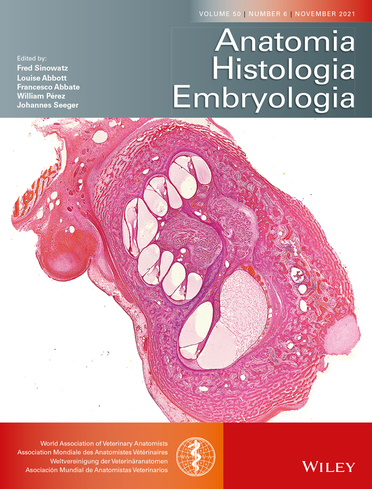
記住我


South American Camelids (SACs) can suffer from locomotor disease (Cashman et al., 1999; Hunter & Semevolos, 2013; Kaneps, 1996), especially of the condition of metacarpo/-tarsophalangeal hyperextension. This pathology involves the interosseous muscle (IOM), which is why this structure has been in the focus of camelid research (Constantinescu, Reed, & Constantinescu, 2008a,2008b; Galotta & Arzone, 2003; Reed, 2009). The palmar/plantar aspect of the distal limb of the four SAC (also called “lamoid”) species – lama (Lama glama, Linnaeus, 1758), alpaca (Vicugna pacos, Linnaeus, 1758), guanaco (Lama guanicoe, Müller, 1776) and vicuna (Vicugna vicugna, Molina, 1782) – has been found to be macroscopically equivalent, but we lack histologic data. The interosseous muscles, appearing in their original fleshy form, for example in primates, carnivores and suidae (König & Liebich, 2019; Lewis, 1965), have undergone adaptive changes in many large grazing animals such as horse, cattle and SACs (Seiferle & Frewein, 2004). In the horse, many studies have focussed on the histological structure of the IOM, which appears mainly collagenous with some muscle fibres and adipose as well as nervous tissue (Bischofsberger et al., 2006; Kaminski, 2006; Nagy & Dyson, 2012; Shikh Alsook et al., 2013; Souza, 2010; Wilson et al., 1991). The IOM of the lama is reported to be mainly tendinous macroscopically (Constantinescu et al., 2008a; Galotta & Arzone, 2003; Schraml et al., 2021), yet diverse in its histologic composition (Reed, 2009). Reed et al. (2007) mention rudimentary muscle fibres in histological samples of the IOM of the lama.
As detailed histological examinations are largely missing, essential questions about the histological composition of the IOM of SACs remain unanswered. Can our macroscopic findings be confirmed histologically? Are muscle fibres a regular component of the lamoid IOM? How much muscle tissue are we to expect and how is it distributed? Are there interspecific differences at a histological level?
It is crucial to have knowledge of the physiological tissue composition of examined structures for the diagnostical application of imaging techniques–essentially to be able to distinguish the healthy from the pathological. In MRI, for example, muscle fibres or adipose tissue embedded in tendinous tissue could otherwise be mistaken for lesions (Bischofsberger et al., 2006).
We hope that this study will contribute to a more complete knowledge and understanding of the nature of the lamoid IOM in the view of biomechanics and evolution and reveal pitfalls when applying diagnostic imaging techniques.
2 MATERIALS AND METHODS 2.1 AnimalsLamas and alpacas were obtained from Peruvian slaughterhouses, guanacos selected from individuals hunted commercially in Chile. In the group of vicuna, only naturally deceased animals were sampled, as this species is protected by law (United Nations, 2019). In the Peruvian highland regions in question, lamas and alpacas are bred extensively. Guanacos and vicunas are feral–only vicunas are typically handled once a year in a shearing procedure. One fore- and one hindlimb of 10 animals per species were collected for histology. For the origin and inclusion criteria of the sampled animals, refer to Table 1.
TABLE 1. Origin and inclusion criteria of sampled animals Species Origin Parameter Inclusion Criteria Alpaca slaughterhouses: Ayaviri and Macusani, Puno, Peru; Chuñuranra, Huancavelica, Peru Breeding/management Extensive Age 2–4 permanent incisor teeth1 Lama slaughterhouses: Ayaviri and Macusani, Puno, Peru; Chuñuranra, Huancavelica, Peru Body condition2 2–3 Reason of death Slaughter for human consumption Guanaco commercial hunt by Frigorífico McLean in San Gregorio, Magallanes, Chile Distal limb No apparent lesions Sample time frame 5 3 hours from slaughter Climatic conditions 5–20 degrees from sampling to processing Vicuna cadavers located in the areas of Nacional Reserve Pampa Galeras Bárbara D’Achille and Community grounds of Lucanas, Ayacucho, Peru. Breeding/management Extensive Age All ages Body condition2 1–2 Reason of death Sarcoptic mange or traffic accidents4 Distal limb Skin lesions acceptable, no apparent pathology on tendons Sample time frame 5 3 hours from encounter Climatic conditions6 1–10 degrees overnight 1Earliest eruption of first incisor pair 2 years and 1 month, latest eruption of second incisor pair 3 years 3 months (Wheeler, 1982). 21: emaciated, 2: thin, 3: optimal (Alpaca Association New Zealand Inc., 2005). 3Verified by autopsy. 4Empirically based assumptions of the author. 5Time from slaughter/locating of corpse until analysis of limb. 6Previous 3 night's temperatures for cadavers found in the countrysid.e. 2.2 Study design and data collectionWe collected the limbs of 40 animals for morphometry and histology. Two limbs of each animal were used for morphometric measurements published in a previous article (Schraml et al., 2021), the others for the histological examination presented in this paper. One limb of a vicuna had to be dismissed. In total, the IOMs of 79 extremities were analysed. Different limbs were used for histology and morphometry because the morphological examination required more time and manipulation of the tendon sections. This could have impaired the sample quality for histology. In general, two ipsilateral left limbs were used for histology, saving cases in which the sample quality of the contralateral limb seemed superior (i.e. because one limb showed subcutaneous hematoma). The IOM of each collected limb was sampled at three defined levels, which are depicted in Figure 1.

Measure–and sample points–schematic drawing and definition. The sample location for the proximal sample point resembles about 15% of the metapodial length of the respective species. As alpacas and vicunas are smaller than lamas and guanacos, there is a difference in the absolute distance of the proximal sample point to the proximal metapodial rim. The location of the proximal and middle sample levels on each limb was determined with the help of a conventional measuring tape. The distal sample point was determined by orientation on the anatomical structures. Abbreviations: DDFT, Deep digital flexor tendon; IOM, Interosseous muscle; Nr°, Number of sample location in technical drawing; SDFT, Superficial digital flexor tendon
The obtained samples were marked for direction with histological marking colours, fixed in 4% buffered formaldehyde solution for a minimum of 7 days and then immersed in a softening solution of 4% phenol in 70% alcohol during another 7 days. A decalcifying solution on the basis of hypochloric [Osteomoll by Merck KgaA] or nitric acid [Solucion Decalcificante by Stainlab] was used, depending on the availability of the chemical, until samples were soft enough to be cut. After dehydration in xylol, the fragments were embedded with paraffin. We then obtained transverse sections with a thickness of 3–6 micrometres with a rotary microtome [RM2125 RTS Leica Microsystems] using high profile blades. Ice and ice water were used to temperate paraffin blocks and facilitate sectioning. After mounting, drying, and hydrating, these sections were stained with a Masson-Goldner Trichrome dye. It stains the nucleus black, muscle fibres brick red and collagen tissue light green (Merck KGaA, 2008). The staining protocol was adapted from the original protocol by Merck as needed. Sections were dehydrated and finished by applying a cover glass with the use of Canada balsam.
2.3 Data analysisFinished slides were digitalized in a histological scanner [Pannoramic Scan II, 3D HISTECH Ltd.], viewed with CaseViewer software [Version 2.3, 3D HISTECH Ldt.] and analysed with the program ImageJ [Rasband, 1997–2020, Version 1.52a]. Slides were disqualified for blurriness or incomplete scanning. A total of 238 slides were found apt to be analysed. For qualitative analysis, samples were first examined as an overview for their external shape and main tissue components as well as tissue distribution. They were then viewed with maximum enlargement, which corresponds to a pixel size of 0.24 µm, to better identify tissue components. The observations were documented and each localization was compared between species. For quantitative analysis, the slide images were further processed in ImageJ. Margins of the sample were obtained using the option “Color Threshold” and improved with manual corrections. Then, muscle tissue was selected manually with a selection tool.
2.4 Statistical analysisData exploration and statistical analysis to address our study questions were carried out with R [Version 1.2.5042]. We considered an effect significant at p < 0.05, with p < 0.001 defined as highly significant. To answer the issue: ‘How much muscle tissue are we to expect and how is it distributed?’ we used the ratio of the area of muscle tissue with the overall cross-sectional area (MTr) as a response variable. Explanatory variables included ‘sample level’ (SL), ‘fore-/hindlimb’ (L) and ‘species’ (SP), employing them as factors. To evaluate the quality of the measurement process, a second operator measured 10 randomly selected samples and we tested the results of MTr of operators A and B for collinearity with two-sided Spearman and Pearson correlation tests. The continuous response variable MTr was visually examined for distribution. This revealed a half-normal data distribution, as no negative values for the response variable exist and values of zero and close to zero are dominating. To underpin qualitative results, the minimum, maximum, mean and standard deviations were calculated for the muscle content of each species overall and for each sample level. We then applied an ANOVA and tested the effects of SL, SP and L explaining MTr. In a subsequent step and based on the ANOVA model, we applied simultaneous inference procedures based on ‘Tukey’ post hoc tests (function ‘glht’) to differentiate for the effect and direction of effect of each factor level, thereby adjusting the p-values to account for multiple testing. We examined all three factors in this manner. This helped clarify if there are differences in the amount of muscle tissue between species, at the different sample levels of the IOM, and on the fore- in comparison to the hindlimb. To verify our statistical approach, we also ran alternative statistic models, which can be found in the Supplementary Material. The obtained results seem robust against different assumptions of the distribution of the response.
3 RESULTS 3.1 Descriptive analysisOn the forelimb, the IOM proximally shows an irregular blunt sickle-form. Here, it has to be separated by dissection from the palmar fascia, with which it intermingles fibres on its lateral side (compare with Schraml et al., 2021, and Figure 3). On the hindlimb, its shape at the same level is more compact and trapezoidal, and it does not present connections of dense collagenous tissue to other structures. The IOM of fore- and hindlimb at all sample levels appears light green, as collagen fascicles dominate the area. A large nerve often shows laterally and can appear alongside the margins or inside the area of the IOM. In two vicunas, we found larger amounts of nervous tissue embedded into the medial side of the IOM at this level. About half of the samples show small amounts of muscle tissue in the medial half close to the dorsal margins, adjacent to the bone. Larger areas of muscle tissue are rare. On occasion, there can be large amounts of adipose tissue infiltrating the IOM from the medio- or the centro-dorsal margin towards the core. Small patches of adipose tissue with little arterioles and venules are often scattered throughout the sample.
At the middle level, larger amounts of muscle fibres are visible, but the vast part of the IOM is still made up of dense connective tissue. The IOM is of blunt trapezoidal shape, with the convex side facing the metapodium. Here, the transition from loose connective tissue to the dense connective tissue of the IOM can be gradual, especially in the centre. Muscle tissue typically forms the outline of a triangle close to the medial margin of the IOM. From there, it is often arranged in lines parallel to the bone-margin. One to two areas of nervous tissue embedded in a small amount of adipose tissue exist centrally, and occasionally, more muscle tissue is present close to these. Adipose tissue occurs alongside all muscle tissue, and the proportions between the two can vary greatly. We note the absence of disperse adipose tissue with vessels as in the proximal part.
The four terminal branches of the IOM macroscopically never separate completely. At the distal sample location, the IOM presents the shape of two bilobed branches with more or less narrow waists. Microscopically, a separation in four oval terminal tendons can be observed in some slides, where the described “waist” is filled with only loose connective tissue. Dorsally, the exact margins of the IOM are also often unclear as there are large amounts of loose connective and adipose tissue with nerves and vessels between the IOM and the adjacent bone margin. The change from loose to dense connective tissue is continuous, especially between the branches of the IIIrd and IVth digit and the fused axial and abaxial branches. Large amounts of muscle tissue, vascular- and nervous tissue can also be embedded within the dense connective tissue of the IOM in these regions. Muscle tissue is mostly present at the point of connection of the axial and abaxial branches and on the interdigital margins.
Overall, the IOM is mostly formed by dense connective tissue with minor patches of muscle and nervous tissue accompanied by adipose tissue and small vessels. A representative image is provided in Figure 2. The IOM appears as a single compact structure; the tissue composition does not follow a symmetrical pattern. At the middle sample level, we can always detect muscle tissue, even though it may only consist of a handful of fibres. The IOM can, in occasions, present disperse adipose tissue at the proximal sample level. The margins between loose connective tissue of the surroundings and dense connective tissue of the IOM are often continuous, especially at the middle and distal sample levels. An overview of the histological aspect of samples is provided in Figure 3.

Tissue components of the South American Camelids’ interosseous muscle–example of an interosseous muscle of the left hindlimb of an alpaca at middle sample level. Abbreviations: A, Adipose tissue; Art, Artefact (specifically colouring and shrinking artefacts, tissue folding); C, Collagen–dense connective tissue; L, Loose connective tissue; M, Muscle tissue; N, Nervous tissue; V, Vessels. This image has been edited with the program Adobe Photoshop Lightroom Classic [Version 8.0, 1193777] to clean up the background and correct its white balance

Overview of different sample levels–examples for variation in the amount of muscle tissue. From top to bottom: proximal, middle and distal sample locations. Left: relatively high amount of muscle fibres. Right: low amount of muscle fibres. Note the generalized colouring artefact in the bottom left section. The slides are examples of the different sample levels of the following South American Camelids: Top left, top right, middle left: Left anterior limb of lama; middle right, bottom right: left posterior limb of lama; bottom left: left anterior limb of alpaca Abbreviations: A, adipose tissue; Art, artefact (specifically colouring and shrinking artefacts, tissue folding); C, collagen–dense connective tissue; Col, colouring (histological colouring to mark for direction); L, loose connective tissue; lat, lateral; M, muscle tissue; med, medial; N, nervous tissue; V, vessels. This image has been edited with the program Adobe Photoshop Lightroom Classic [Version 8.0, 1193777] to clean up the background and correct the white balance
3.2 Morphometric analysisCorrelation between operators in the quantitative measuring process of MTr is shown to be high. We obtained rho-values >95% from correlation tests (0.952 Spearman rank correlation test, significance level <2.2e-16, 0.988 Pearson's correlation coefficient, significance level 8.27e-08). To quantify the results from the descriptive analysis, we then measure the muscle tissue content of the IOM. It varies from 0 to 19% with a mean of 1.7%. This clearly shows that muscle tissue is not the main component of the lamoid IOM. The mean of MTr was 1.7 ± 2.8%, the median 0.8%. Results are presented in Table 2.
TABLE 2. Area of muscle tissue occupying the total cross-sectional area of the IOM in per cent Species All species Alpaca Lama Vicuna GuanacoRatio muscle tissue/total cross-sectional area
Min 0.00 0.00 0.00 0.00 0.00 Max 19.3 19.3 7.9 4.8 4.9 Mean ± SD 1.7 ± 2.8 3.3 ± 4.8 1.8 ± 1.9 0.9 ± 4.8 0.8 ± 0.9 Nr° NA 14 1 0 5 8 Abbreviations: Max, Maximum; Min, Minimum; Nr° NA, Number of unavailable values within the sections. Slides were disqualified (= NA) because of blurriness of the scanned image or incomplete scanning; SD, Standard deviation.Mean muscle fibre content at medium sample level is 3%, with a minimum of 0.4 and a maximum of 15.2%, confirming the observations from the descriptive analysis.
After proofing the results from the qualitative analysis, we checked for the influence of the factors SL, L and SP on MTr. The ANOVA demonstrated the influence of the factors on MTr by rendering highly significant results for the factors SL and SP (p < 0.001), as well as significant results for L (p < 0.05). The corresponding F-values are shown in Figure 4.

F-values of the factors level, species and limb on the amount of muscle tissue within the interosseous muscle. Abbreviations: n, Number of sampled animals. The significance level is indicated by asterisks: 0 ‘***’ 0.001 ‘**’ 0.01 ‘*
Taking a closer look at each factor, Tukey post hoc tests helped to reveal the size and direction of effect for each factor level. We found for SL that significantly more muscle tissue is present at the middle sample level than in the proximal and distal areas of the IOM. However, the difference between the proximal and distal sample levels was not significant and visualizations hint that the proximal sample level could contain even less muscle fibres than the distal sample level. For graphical results of the significance of sample levels, see Figure 5. In regard to the factor SP, our analyses demonstrate that alpacas have a significantly higher MTr (p-values ≤0.0052) than all other species. Lamas are ranked 2nd as they present significantly more muscle tissue than guanacos (p < 0.05, t-value =2.7), whilst there are no significant differences between lama and vicuna or vicuna and guanaco. Results are summarized graphically in Figure 6.

Muscle tissue ratio of the interosseous muscle: Visualization in relation to sample localization and results of Tukey post hoc tests. Note: Number of sampled individuals n = 80. Abbreviations: d, Distal sample level; m, Middle sample level; p, Proximal sample level. The significance level is indicated by asterisks: 0 ‘***’ 0.1 ‘ -’

Muscle tissue ratio of the interosseous muscle: Visualization in relation to species and results of Tukey post hoc tests. Abbreviations: A, Alpaca; G, Guanaco; L, Lama; V, Vicuna. The significance level is indicated by asterisks: 0 ‘***’ 0.001 ‘**’ 0.01 ‘*’
For the factor limb, we showed that the posterior limbs show a significantly higher MTr than the anterior limbs (p < 0.05) (Figure 7).

Muscle tissue ratio of the interosseous muscle: Visualization in relation to the factor fore-/hindlimb and results of Tukey post hoc tests. Abbreviations: ant, Anterior; post, Posterior. The significance level is indicated by asterisks: 0.01 ‘*’
4 DISCUSSIONIn the following, we will first discuss the critical points of the methodology of this study to then further cast light on the importance of the results.
As to the animals used for this study, inclusion criteria for sampled alpaca, lama and guanaco were narrow in relation to age, management and cause of death, to minimize the chance of lesions. For vicuna, we could not provide similarly narrow criteria because of them being considered an endangered species. As a result, they were older on average, as well as chronically sick.
Due to general management practice, it was not possible to obtain an individual history of each animal or a lameness exam prior to slaughtering. For further studies, collecting this kind of information for each animal would be ideal.
Various problems occurred during histologic processing, of which some were due to our study design. Shrinking artefacts are a common problem associated with tissue fixation and processing. They typically occur when formaldehyde is used as a fixative, especially in combination with paraffin as an embedding medium. Due to complicated time management, some samples remained in formalin for prolonged periods. This provokes the formation of stable crosslinks between macromolecules (Suvarna et al., 2013), which may have further accentuated tissue shrinking and toughness of the samples.
Histologic processing was difficult due to the mentioned hardness of the collagenous tissue, and we recommend cryosection for coming studies as previously proposed for the horse (Shikh Alsook et al., 2013). The use of phenol as a softening agent (Nagy & Dyson, 2012; Schramme et al., 2012; Shikh Alsook et al., 2013; Wynnchuk, 1992) in combination with decalcifying solutions made sectioning possible, but not at high quality. Even though we obtained very good colour contrast with Masson-Goldner Trichrome stains in pre-trials, uneven thickness and colouring artefacts of samples impaired the contrast to a point where an automatic assessment of tissue proportions was not possible in the main study and evaluation had to be carried out manually.
Problems with the covering medium on occasions led to incorrect mounting and, therefore, blurriness of the image from the histological scanner, in a way that some slides had to be dismissed after digitalization. Most NAs occurred due to such scanning problems. As the resulting NAs are unevenly distributed (view Table 2) between species, sample size suffered especially for guanaco, which could bias the results.
The tissue processing, colouring and mounting procedure should be modified for further studies to obtain optimal results.
Our approach to the data analysis is especially important to keep in mind when interpreting the results, as there are numerous studies in horses using very different proceedings. An analysis of MTr on digitalized slides with the software ImageJ has been carried out by Shikh Alsook et al. (2013) in equines. They document tissue ratios with the total CSA of the IOM. We found this to be a convenient and reproducible method of determining tissue components.
The high interoperator correlation demonstrates the quality of our measurement process. It confirms that even though staining results were sometimes poor, discrimination between muscular tissue and other tissue components was possible with high accuracy and that the overall tissue area was easy to determine. Errors from influences like tissue shrinking have to be taken into account separately, as they equally impact both operators.
The statistical analysis has some limitations. As described in the ‘materials and methods’ section, our data showed a non-normal distribution and sample sizes were relatively small. Even though we employed different statistical models and obtained robust results, the distribution may impair the value of the statistic tests. Further studies with larger sample sizes are recommended to verify the tendencies observed in this investigation.
Our main results show the SAC IOM as a macroscopically and histologically complex and unique structure. The IIIrd and IVth interosseous muscles in lamas, alpacas, guanacos and vicunas form a single structure without perceivable septa at the proximal level. In the horse, in contrast, this part is often described as showing two lobes (Geburek & Wissdorf, 2014; Nagy & Dyson, 2012; Schulze, 2007).
The SACs’ IOM is largely tendinous. This can be seen as an adaptation to grazing life in more or less plain environments (Franklin, 1982, cited by Lamo, 2011). In the horse, it has been shown that the tendinous structures of the fetlock are the secret to long-term indefatigable load bearing on a hyperextended fetlock, as well as elastic energy-saving and impact-absorbing mechanisms at stride (Hildebrand & Goslow, 2004; Wilson, McGuigan, Su, & Bogert, 2001). One may assume that the less muscular and the more tendinous the IOM, the more apt it is to the biomechanical challenges of the described lifestyle. The collagenization may have advanced further in the larger of the wild species - the guanaco of the Patagonian plains - than in the lighter, highland vicuna. In equines, Schulze (2007) demonstrated that there are more muscle fibres in the IOM of ponies than large horses. Additionally, the lama, domesticated from the guanaco with some hybridization with the alpaca (Kadwell et al., 2001), has been selectively bred by Precolumbian cultures for loadbearing (Wheeler, 1995), which could have influenced the composition of the IOM. The morphometric analysis of the histologic sections brought about interesting results. Not surprisingly, muscle tissue content was significantly higher at the middle level than at the proximal and distal sample levels. This level resembles the evolutionary body of the interosseous muscle, which is why we expect remnants of muscle tissue especially in this area. We did, however, not find a significant difference between proximal and distal sample levels. It was surprising that in many individuals muscle tissue was detectable at the distal sample levels, which coincides with the IOM’s branches. This stands in opposition to the findings on the horse (Mülling, 2015; Shikh Alsook et al., 2013). We should, therefore, not assume properties of real tendons even for the terminal branches of the lamoid IOM. Kaminski (2006) proposes that muscle tissue within the IOM of horses may not be mere remnants, but act as functional tissue in tension control. It is also important to mention that the maximum value of MTr of all samples occurred at the distal sample level. As this value was an outlier from the dataset of the distal sample level, an alternative explanation seems suitable: The shape of the sample that renders this high value suggests it might have been cut more proximally than usual. This suggests an increase of MTr between middle and distal sample level that we did not detect because of level spacing. Subsequent investigations should aim to process more levels per IOM to get continuous data for muscle fibre distribution. Additionally, further studies are needed to understand the role of muscle fibres within different parts of the lamoid IOM. It is important to know if they are just leftovers from their evolutionary origin or play a functional role. As most studies of the horse either assess only part of the IOM (Bischofsberger et al., 2006; Nagy & Dyson, 2012), do not give absolute numbers (Shikh Alsook et al., 2013) or use measuring techniques that are not comparable to ours (Kaminski, 2006), a quantitative comparison of muscle tissue content is difficult. Nagy and Dyson (2012) cite unpublished data of Dyson et al., proposing that muscle tissue occupies between 2% and 11% of the IOM in horses without going into more detail. Shikh Alsook et al. (2013) show means between about 6.5% and 9% in their figures, depending on the horses’ age, and Bischofsberger et al. (2006) mention means of about 13.0% ± 6.7% at a localization that approximately coincides with our proximal sample level. Viewing these numbers, they seem to hint that SACs show an even smaller ratio of muscle tissue in their IOM than horses.
We saw that the factor species has a significant impact on the proportion of muscle fibres. While we found significantly higher percentages of muscle tissue in alpacas than in any of the other species, differences between lama and vicuna, for example, were not statistically significant. Vicuna are the smallest of the SAC species, followed by alpaca (Ccora Esteban & Condori Enriquez, 2019; Grund, 2014; Lamo, 2011). The much larger guanaco and lama resemble each other approximately in size and weight (Franklin et al., 1997; Lamo, 2011; Smith et al., 1992). These apparently similar pairs of species do not show the expected parallels in the MTr of their IOMs, which dismisses our theory that lighter weight and smaller size correlate with higher muscle tissue content of the IOM. It seems to indicate that other factors, as domestication or lifestyle/training, have an impact on tissue composition. Both guanaco and vicuna possibly move and run more than their domesticated colleagues. Guanacos in the Patagonian plains additionally frequently jump high sheep wire fences (Rey et al., 2012), presumably putting peak strains on the IOM of their forelegs. This exercise exceeds the training the other species undergo in daily life and could possibly add to an altered tissue composition with a more tendinous IOM. In the case of vicuna, their on average older age and maybe also chronic sickness could be an additional factor for low MTr. Various authors have claimed the muscle content of the IOM to diminish with age in different species: Ruminants in general (Seiferle & Frewein, 2004), cattle (Müller-Calgan, 1954, cited by Nickel et al., 2004; Nuss, 2017), ovine (Köppen, 2014; Mascarello & Rowlerson, 1995) and the horse (Seiferle & Frewein, 2004; Shikh Alsook et al., 2013). We should, therefore, consider that their higher average age could erroneously lower the values of MTr in vicuna.
This study shows that the IOM in SACs has undergone a transformation to a largely tendinous structure and that a large individual variation of tissue proportions within the IOM seems to be normal. A statistically significant difference in muscle content between species could be shown, with alpacas showing significantly more muscle tissue within the total cross-sectional area of the IOM than any of the other camelid species. A comparison with the horse reveals some important parallels in histologic composition. However, different to the horse, domestic lamoids present relevant amounts of muscle tissue in the distal parts of the IOM, of which the biomechanical purpose still remains unclear.
ACKNOWLEDGEMENTSWe very much appreciate the incountable contributions made by Dr. Jenny Hagen. The team is obliged to Dr. Karsten Winter for making histological scanning possible even during a pandemic. Also, the authors want to genuinely thank Miluska Beatriz Navarrette Zamora for all her help at the UNMSM. We are grateful to Jose Angel Sarmiento with the whole community of Lucanas as well as the staff of the National Reserve of Pampa Galeras Barbara D’Achille for their hands-on help with the complicated sample acquisition of vicuna material. Special thanks to Liber Lazo with the staff of Frigorífico MacLean for the effective and at the same time very enjoyable time in Patagonia.
留言 (0)