
記住我

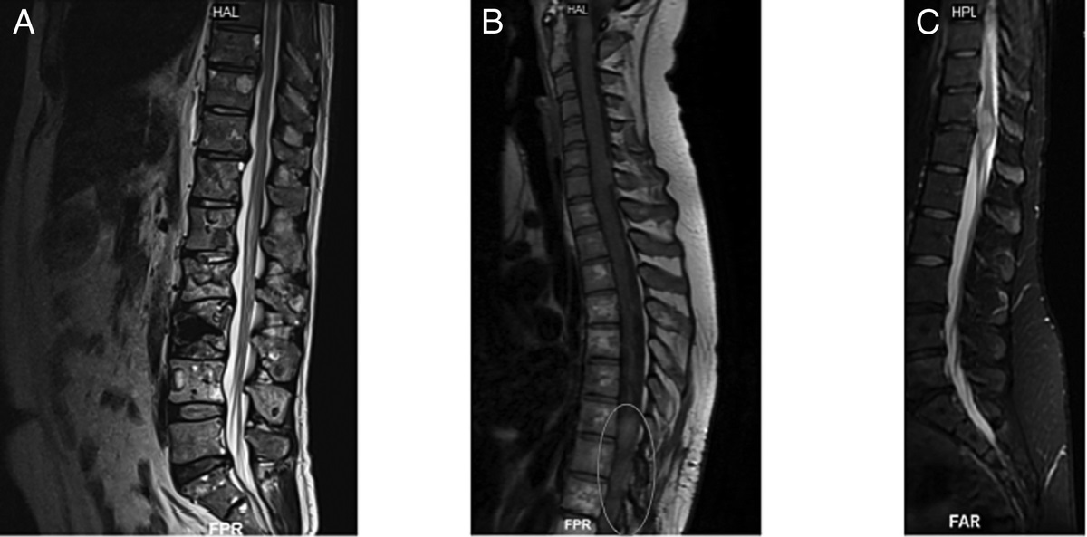


A 35-yr-old man presented to our emergency department with traumatic, central, nonradiating, 5/10 low back pain after a fall. His medical history was unremarkable. His physical examination was normal. A lumbosacral computed tomography (CT) scan was performed because of the history of trauma. Computed tomography examination has a higher sensitivity and specificity for evaluating spine injury compared with plain film radiographs.1 Sagittal view of CT (Fig. 1) showed incidental finding of an intervertebral disc at S1-2 level. There was no fracture. The patient’s pain was due to muscle spasm, and he was treated with nonsteroidal anti-inflammatory drugs. His pain completely resolved in the emergency department, and he had no complaints afterward.
 FIGURE 1:
 Sagittal image of CT of 35-yr-old man. There was a residual intervebral disc (arrows) at S1-2 level.
The differential diagnosis of a residual intervertebral disc on CT scan includes tarlov cysts, spinal synocial cysts, meningoceles, nerve sheath tumors, and spinal metastases.2 Variations in sacral morphology are common, and residual intervertebral discs are a normal anatomic variant. Persistence of an intervertebral discs at the sacrum is an interesting finding of which clinicians should be aware. Anatomic variations should be taken into consideration when diagnosing and treating sacrum-related diseases.
The sacrum is a large, triangular bone constituted by the fusion of five sacral vertebrae. The intervertebral discs are frequently absent in the sacrum and coccyx because of the fusion of these vertebrae. The five sacral vertebrae are connected by intervertebral discs during childhood with the subsequent fusion of the S4-5 and S3-4 levels in late adolescence and fusion of the S2-3 and S1-2 levels by the third decade of life. It is not infrequent to detect a residual disc at S1-2 levels on studies, especially in young adults. Satheesha Nayak et al.3 reported that there was presence of residual intervertebral disc between S1 and S2 in 39% of the pelvises on cadaveric study. An expansive radiological study needs to be conducted to get a better understanding of the anatomy of this region. It is important that clinicians recognize that the presence of a sacral intervertebral disc is a normal anatomical variant that should not be mistaken for a pathological condition.
LEARNING POINTS Presence of intervertebral discs in the sacrum is a normal anatomical variation, and knowledge regarding its prevalence may be helpful. Satheesha Nayak et al.3 reported that there was presence of residual intervertebral disc between S1 and S2 in 39% of the pelvises on cadaveric study. Variations in sacral morphology are common, and these anatomic variations should be taken into consideration when diagnosing and treating sacrum-related diseases. Examples of other sacral anatomic variations are angel-wing sacrum, accessory sacroiliac joints, complete agenesis of the dorsal wall of the sacral canal, developmental defects in the ala of the sacrum, developmental absence of a portion of the right ala of S1, sacral ribs, etc. Images of residual intervertebral discs can be mistaken for pathological conditions, especially tarlov cysts and spinal synocial cysts. Spinal synovial cysts are cystic formations connected to the facet joint and containing synovial fluid. Tarlov cysts are cerebrospinal fluid-filled dilatations of the nerve root sheath at the dorsal root ganglion. REFERENCES 1. Parizel PM, van der Zijden T, Gaudino S, et al.: Trauma of the spine and spinal cord: imaging strategies. Eur Spine J 2010;19(suppl 1):S8–17 2. Smith AB, Soderlund KA, Rushing EJ, et al.: Radiologic-pathologic correlation of pediatric and adolescent spinal neoplasms: part 1, intramedullary spinal neoplasms. AJR Am J Roentgenol 2012;198:34–43 3. Satheesha Nayak B, Ashwini Aithal P, Kumar N, et al.: High incidence of persistence of sacral and coccygeal intervertebral discs in South Indians—a cadaveric study. J Can Chiropr Assoc 2016;60:182–9
留言 (0)