This case proved to be a diagnostic challenge. The diagnosis of sympathetic ophthalmia was reinforced by the positive response to immunosuppression alone without adjunctive antimicrobials or ATT. There were no episodes of reactivation throughout the 1 year that he was on immunosuppression alone. A differential diagnosis of ocular tuberculosis should be considered due to its endemicity in this region, its ability to present similarly, and the positive TST in this patient. However, ocular tuberculosis would have worsened with immunosuppression alone and without the appropriate administration of ATT [11]. This patient showed complete resolution of ocular findings without any recurrence.
Opportunistic infections are the commonest cause of lung changes in individuals with immunosuppression [12]. However, in the absence of a causative organism and no significant improvement with broad-spectrum antibiotic therapy, the differential diagnosis of drug-induced toxicity has to be considered. In our patient, the diagnosis of azathioprine-induced lung injury was entertained as all infective workup were negative, and he did not improve with a course of broad-spectrum antibiotics. He did not exhibit any of the common side-effects of azathioprine which include bone marrow suppression, nausea, vomiting, anorexia, hepatotoxicity and hypersensitivity reactions [13]. Azathioprine-induced lung injury is a rare dose-dependent adverse effect. It should be considered in patients on immunosuppression who present with fever, acute respiratory symptoms and hypoxia due to pneumonitis, and acute respiratory distress syndrome (ARDS) [12,13,14]. CT thorax in these patients shows nodular hyperdensities with ground-glass areas without pleural effusion or focal lung lesions [12, 14]. Clinical improvement usually occurs rapidly within 1 to 2 weeks of discontinuation of azathioprine [12,13,14]. In our case, the patient’s symptoms were insidious. He was afebrile, not hypoxic, and his CT thorax did not show any ground-glass appearance typically seen in azathioprine-induced toxicity. There was also no improvement despite azathioprine cessation. At this point, an alternate diagnosis was considered.
Due to his risk factors of immunosuppression, cigarette smoking and significant history of tuberculosis exposure, the possibility of pleural tuberculosis was considered. The insidious nature of his symptoms combined with the significant weight loss, CT thorax findings and presence of an exudative pleural effusion supported the diagnosis. However, the absence of MTB yield from any body fluid was a dilemma.
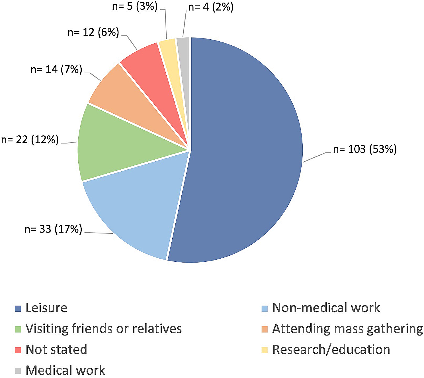
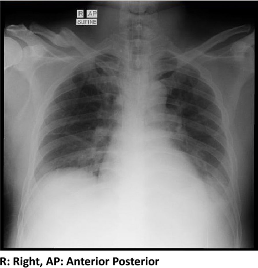
Our patient had a right-sided small pleural effusion. TPE are usually unilateral and can be of any size. A case series of 333 patients showed 86.5% having unilateral pleural effusions and 20.4% having small pleural effusions [15].
The CT thorax, in this case, revealed more findings compared to the initial chest radiograph. This is further supported by Zhai et al., who stated that CT thorax is more sensitive than chest radiography in detecting lung parenchymal changes in TPE, with approximately 40–85% of patients demonstrating parenchymal disease [15]. The CT thorax findings of pleural effusion, subpleural nodule, tree-in-bud changes, lung parenchymal consolidation and mediastinal lymphadenopathy can be found in tuberculosis [9, 16].
A predominantly neutrophilic TPE indicates an earlier MTB infection phase and a higher MTB yield than a predominantly lymphocytic TPE [17]. This would explain the presence of solely mononuclear leukocytes in our patient’s pleural fluid, as he already had the symptoms for a total of 3 weeks before sampling of the effusion. Unfortunately, it was not specified whether these mononuclear leukocytes were lymphocytes or monocytes. The absence of MTB yield in our patient is expected, given the longer illness duration.
TPE is paucibacillary, as it is a hypersensitivity reaction to mycobacterial proteins rather than frank infection [18]. This may also explain the lack of MTB detection from our patient’s pleural fluid analysis. Mycobacterial cultures from pleural fluid have a higher sensitivity for AFB (24–58%) than direct smears (< 5%) as direct smears would require a greater bacilli concentration of 10,000/mL compared to 10–100/mL required for culture [19, 20]. GeneXpert, a type of nucleic acid amplification test (NAAT), utilizes a polymerase chain reaction (PCR) method to detect small amounts of MTB genetic material in samples [9]. It has a pooled sensitivity of 50.9% against culture from pleural fluid [21]. However, pleural biopsy has the highest sensitivity in diagnosing pleural tuberculosis with either the presence of granulomas or detection of MTB from direct smear, cultures or NAAT (46–88%) [7, 8, 10, 19, 22]. Although a pleural biopsy may have provided a positive result, the positive clinical and radiological response to ATT, demonstrated in this case, is sufficient to make the diagnosis of pleural tuberculosis [22].
The decision to treat LTBI with ATT is multifactorial depending on the TB burden and economic status of the country, and risk stratification of an individual for tuberculosis activation [23]. According to the World Health Organization’s recommendation for screening and treatment of LTBI, corticosteroid administration and cigarette smoking alone confer a low risk for tuberculosis activation and there is no conclusive evidence of benefit from ATT prophylaxis in these groups of patients [23, 24]. However, the addition of azathioprine may increase this risk and hence, treatment of LTBI in an individual with multiple risk factors should be considered. Additionally, vigilance for reactivation of tuberculosis is required in patients with LTBI on immunosuppression.


留言 (0)