
記住我


Nicolas Eftychioua, Jaime Veraa, Henryk Barthelb, Matti Shuererb, Michael Rullmanb, Osama Sabrib, Ana Andradec, Johan Lijlad, Helena McMeekine, Mark Aplina, Sabina Dizdarevica,c
aBrighton and Sussex University Hospitals, Brighton, United Kingdom.
bUniversity of Leipzig, Leipzig, Germany.
cClinical Imaging Science Centre, Brighton, United Kingdom.
dHermes Medical Solutions, Stockholm, Sweden.
eHermes Medical Solutions, London, United Kingdom
Aim: To assess value of [18F]florbetaben (FBB) in people living with HIV (PLWH) and subjective cognitive impairment (sCI), when HIV associated neurodegenerative disorders (HAND) and other types of dementia are considered as differential diagnoses, and its impact on management.
Method: FFB-PET/CT imaging was prospectively performed in 20(Male=18) PLWH with sCI [median age 59 (13)]. Objective cognitive impairment was defined using the Frascati criteria (fCI). Images were visually assessed by 2 different imaging centres. Semi-quantitative analysis was performed using Hermes BRASS Florbetaben software and by obtaining a composite cortical to cerebellar cortex standardised uptake value ratios (SUVRs) using cerebellum as reference region [SUVR>1.35 (>mv+2SD of healthy controls)]. Clinician diagnostic confidence before and after (scale 1 to 10, 1=no confidence and 10=complete confidence) was assessed.
Results: Fourteen/20 PLWH (70%) had objective fCI; in total, 4/14 (29%) had visually and semi-quantitatively positive scan; 2/4 (50%) were clinically diagnosed with AD type dementia; 2/4 (50%) had mild CI and remained under close clinical follow up. Six/20 (30%) had no fCI and negative FFB scan.
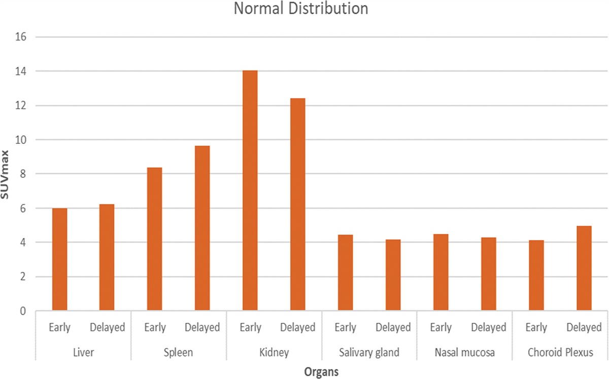
Regionally the greatest SUVRs were observed in the posterior cingulate and superior temporal and frontal superior lobe.
FFB significantly increased clinical confidence in all 14 fCI but did not change in 6 neg fCI patients. (Mean 6.9 to 8.1; p<0.005)
Conclusion: FFB can identify amyloid plaque deposition in PLWH. It increased clinical confidence in 70% of cases. Negative scan is reassuring for AD type dementia. FFB role in differentiating HAND from AD type dementia needs to be established.
2. Interobserver Variability in the Qualitative and Quantitative Analysis of Cardiac MIBG Scintigraphy for the Diagnosis of Lewy Body DisordersRob Foleya, Chris Greena, Isabel Laurenceb, Stewart Redmana, Richard Grahama, David Littlea
aRoyal United Hospital, Bath, United Kingdom.
bSouthmead Hospital, North Bristol NHS Foundation Trust, Bristol, United Kingdom
Introduction: [123I]-MIBG cardiac scintigraphy is a tool to detect cardiac sympathetic denervation, which can be helpful to differentiate between different Parkinsonian syndromes. Images can be assessed visually and quantitatively. The aim of this study was to review cardiac MIBG studies at 2 nuclear medicine departments.
Methods: Planar images were acquired in the anterior view at 15 minutes and 4 hours using a double-headed gamma camera (GE Healthcare) and a low-energy collimator. The heart: mediastinal ratio (HMR) was measured by 6 different scorers including radiologists and technologists, we also subsequently recalculated HMRs for a medium-energy collimator, using a published formula. The visual and final interpretations (normal, abnormal or borderline) were recorded in each patient.
Results: The cohort consisted of 10 patients. On visual interpretation only, there was a near-perfect agreement between 2 consultant’s interpretation of the scan, κ = .82, (95% CI, .510 to 1.00), p<0.01. However, the final interpretation, with the addition of low-energy HMR, led to fair agreement, κ = .37, (95% CI, .023 to .727), p=0.06. Conversion to a medium-energy HMR led to a significant increase in mean HMR, (1.79 vs 1.36, p=0.02) and when utilised in reporting, resulted in perfect agreement, κ = 1.0, p<0.01.
Conclusion: Agreement on the visual interpretation of cardiac MIBG is near-perfect, however the use of quantified heart: mediastinal ratios can lead to important differences in the final report. After correcting for the use of a low-energy collimator there was perfect agreement in the interpretation of these studies amongst 2 consultant radiologists.
3. Quantification for DaTSCAN: can it change your reports and add confidence?Alp Notghi, Mohamed El-Sayed, Terence Jones, Joseph O’Brien
Sandwell & West Birmingham Hospitals NHS Trust, Birmingham, United Kingdom
Aim: Evaluate to what extent quantification can change DaTSCAN reporting.
Method: 35 consecutive DaTSCAN™ studies were selected. These were acquired on either GE Infinia or Discovery 630 cameras, 3.5 hours after [123I]Ioflupane injection. Images were processed using recommended parameters (OSEM, Butterworth filter critical frequency 0.6, power-factor 10). Specific Binding Ratio (SBR) was calculated using DaTQUANT™ software package (GE Healthcare). The randomised scans were reviewed by three reporters (R1=consultant, R2=registrar, R3=experienced physicist) twice; with and without quantification.
Reporters had to choose: A) Scan report (1:normal, 2:PD/DLB, 3:striatal infarct, 4:technical problem, 5:don’t know). B) Reporting confidence (1 to 5). Wilcoxon Rank test was used for paired matched data, and ANOVA for multiple related data. Chi-square test was used to compare between reporters.
Result: There was a significant difference in report with and without quantification (p<0.001). 20/105 reports changed following quantification (4, 11, 5 by R1, R2, R3 respectively). Reporting confidence increased significantly (p<0.001). There was significant difference between reporters with least change in reports by consultant (p=0.25), then experienced physicist (p=0.031) and most changes by junior reporter (p=0.005). Interestingly, the reporting confidence of the two clinicians was higher with or without quantification than the experienced physicist (p<0.001). The increase in reporting confidence with quantification only occurred with clinicians but remained the same with the physicist. Reporting confidence in cases where quantification affected the report was significantly lower than in cases where the report did not change.
Conclusion: Quantification has significant positive effect on the DaTSCAN interpretation and reporting confidence.
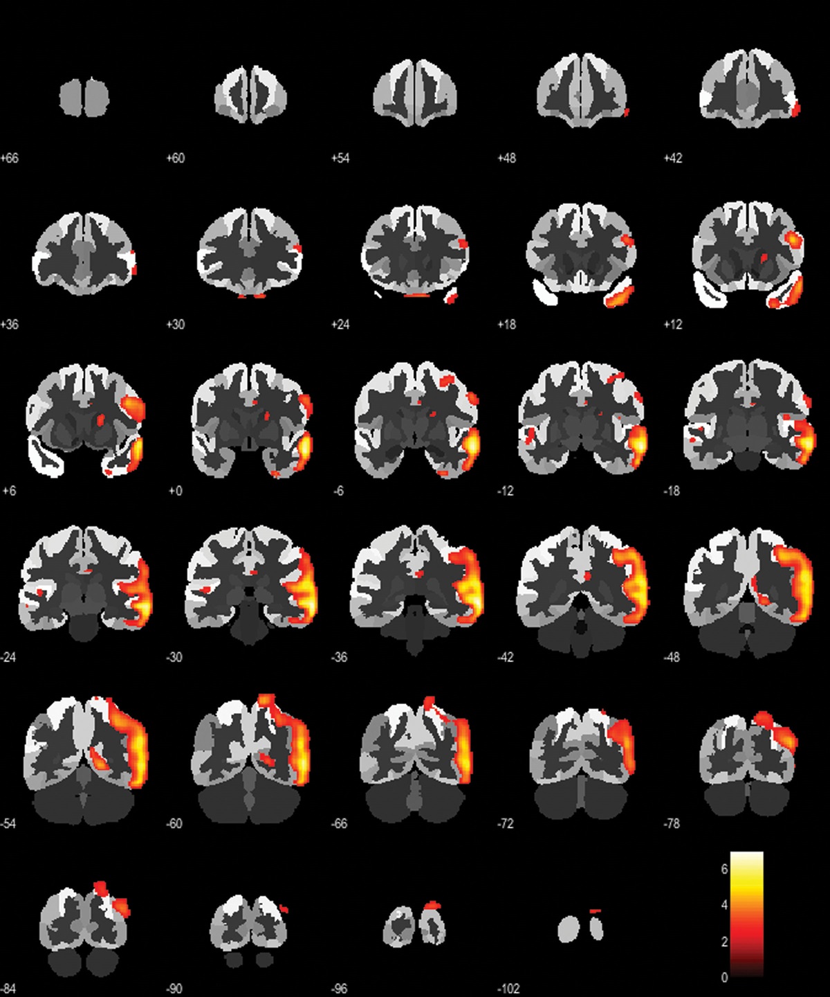
4. Improvement of [18F]FDG PET/CT and MRI concordance in Temporal Lobe Epilepsy Pre-surgical Assessment using Statistical Parametric Mapping Z-scores: 2nd Place Student PrizeMarie Kershawa, Lu Yipingb, Vijay Sawlania, Hayaka Amadac, Simon Hughesd
aUniversity of Birmingham, Birmingham, United Kingdom.
bFudan University, Shanghai, China.
cCity Hospital, Birmingham, United Kingdom.
dUniversity Hospitals Birmingham, Birmingham, United Kingdom
Objectives: This study evaluates the diagnostic performance of statistical parametric mapping (SPM) analysis of [18F]FDG PET/CT in temporal lobe epilepsy (TLE). Our aim is to increase consistency in image reporting and increase confidence in surgical evaluation.
Methods: Thirty-eight patients with TLE who underwent MRI and [18F]FDG PET/CT imaging at the Queen Elizabeth Hospital Birmingham were included. Images were interpreted by visual assessment by radiologists. Quantitative analysis for [18F]FDG PET/CT was performed using SPM. Statistical analyses performed include Kruskal-Wallis and Game Howell tests, analysis of ROC curve and Cohen’s κ statistics.
Results: The standardised uptake value (SUV) ratio for left temporal epilepsy, non-epilepsy and right temporal epilepsy were -1.06±1.01, 0.08±0.79, 0.39±0.73 respectively, exhibiting significant difference between the three groups (p<0.01). In the left/non-left group, the area under curve (AUC) was 0.881 while the cut-off value to separate left temporal epilepsy from non-epilepsy and right temporal epilepsy was -0.305 with 89.7% sensitivity and 88.9% specificity. In the right/non-right group, the AUC was 0.792 while the cut-off value to separate left temporal epilepsy from non-epilepsy and right temporal epilepsy was 0.19 with 83.3% sensitivity and 81.3% specificity. When patients were divided into three groups based on SUV ratio (left temporal epilepsy, non-epilepsy and right temporal epilepsy), there was good inter-method agreement between MRI and SUV ratio (κ = 0.63, 95% CI, 0.42–0.85).
Conclusion: These results indicate that PET/CT-based SUV ratios may have promising diagnostic value in future clinical practice.
5. Approaches of PET metallomics for the study of metal trafficking in vivoGeorge Firtha, Zilin Yua, Joanna Bartnickaa, Fahad Al-salameea, Hannah Greenwooda, Jana Kima, David Parkerb, Philip Blowera
aKing’s College London, London, United Kingdom.
bUniversity of Birmingham, Birmingham, United Kingdom
Essential trace metals such as copper, zinc and manganese play crucial roles in the human body. A disruption to the homeostasis of these metals is implicated in several diseases, most notably prostate cancer, Alzheimer’s disease and diabetes. There is an unmet need to non-invasively study metal trafficking in vivo, with PET metallomics emerging to address this need. We have access to an ever expanding range of radiometals with different imaging properties and relevance to different diseases. The in vivo distribution of 52Mn (t1/2 = 5.6 d; β+ = 30%), 62Zn (t1/2 = 9.3 h; β+ = 3%) and 64Cu (t1/2 = 12.7 h; β+ = 18%) was investigated in healthy BALB/c mice over a day. All radiometals were rapidly cleared from the blood and localised to major organs such as the liver, kidneys and intestines. Interestingly, 52Mn and 62Zn demonstrated significantly higher uptake in the pancreas compared to 64Cu. Although 62Zn has previously been considered a poor choice for PET imaging due to its unfavourable decay to 62Cu (t1/2 = 10 min; β+ = 97 %), our data suggests that 62Zn administered as a citrate complex is handled in vivo primarily as zinc citrate and not copper citrate. Tracking 52Mn and 62Zn in diabetes, cancer and dementia in mice and later in humans will be important
areas for future research. Together these radiometals set the foundation for the study of essential metals in clinical diseases where metal trafficking is disrupted.
6. Maximum acceptable delay in processing [99mTc] Tc-DTPA GFR plasma samplesLaura Perrya, David Woodhouseb, Michael Woodwarda, Zarni Winb, Kuldip S Nijrana
aRadiological Sciences Unit, Imperial College Healthcare NHS Trust, London, United Kingdom.
bNuclear Medicine Department, Imperial College Healthcare NHS Trust, London, United Kingdom
Introduction: Plasma samples for GFR studies must be counted before radioactive decay reduces activity concentration below detectable limits. Since moving from [51Cr] Cr-EDTA to [99mTc]Tc-DTPA for GFR studies the maximum delay between injection and sample counting has decreased due to the difference in physical half-life. The risk of non-diagnostic studies due to equipment downtime for faults and planned maintenance is increased.
This work aimed to establish the maximum delay possible between administration and sample counting which maintains accuracy of the GFR result.
Method: GFR was performed using 10 MBq [99mTc] Tc-DTPA with blood samples taken at 2 and 4, 6 or 24 hours post administration. 1ml plasma samples were counted using a Perkin Elmer 2480 Wizard gamma counter for 10kcts or 45mins. GFR was calculated using the Slope-Intercept method (Fleming JS et al. 2004, 25(8):759-69. Nucl.Med.Commun.). The plasma samples were repeatedly re-counted up to 86 hours post administration. GFR values were re-calculated and compared to the original value.
Results: The plasma samples from 76 patients were included with a mean 4.8 (range 2-10) re-calculations per patient. A total of 364 GFR results were analysed. The change in GFR exceeded ±5% for all samples counted beyond 56hours post administration. The change in GFR was less than ±5% for 99.1% samples counted within 37hours post administration.
Conclusion: Clinically we have implemented a maximum acceptable delay in processing of GFR samples of 37 hours. Our local guidelines require all counting to be completed by 5pm on the day after the patient was injected.
7. Can patient age be used to decide the optimum sample time for single sample GFR studies?Alexander Smout, James Scuffham
Royal Surrey County Hospital, Guildford, United Kingdom
The 2018 BNMS GFR guidelines require the expected GFR to determine the optimum sampling time. McMeekin et al (BNMS2019) discussed that this is chosen as a balance of accuracy and precision of single-sample GFR results (SS-GFR) versus multi-sample results.
This presents logistical issues, leading some centres to explore a fixed sampling time of 4 hours for all patients. We performed analysis on 16203 of our patients’ data with samples at 2, 3 and 4 hours to determine whether the optimum sampling time could be chosen by patient age. The accuracy and precision of our calculated SS-GFR results using an age-based threshold approach were compared to a fixed sampling time of 4h and also to the BNMS2018 eGFR-based approach.
The fixed sampling time of 4h has reasonable precision and accuracy for GFR 40-80 ml/min, above which the precision and accuracy progressively worsen. Performing 2 hours sampling for patients under age 70 and 4 hours sampling for older patients gave the same accuracy as the BNMS guidelines from GFR 50-150ml/min, but worse precision across this range. Interestingly, an age-based method with threshold of age 60 was significantly more accurate than the fixed 4 hours for-all method, and had equivalent precision.
The BNMS guideline method for optimum sampling time had better overall precision and accuracy than either method.
We propose that a reasonable protocol for deciding the optimum sampling time of non-oedematous patients would be to use the eGFR where available (following BNMS guidelines), otherwise taking a 2 hours sample for patients under age 60 and a 4 hours sample for older patients.
8. A feasibility study for half time [99mTc]Tc-MDP whole body bone imagingEmily Fittock, Clare Beadsmoore, Andoni Toms, Ramona-Rita Sultana, David Newman, Davina Pawaroo, Matthew Gray
The Norfolk and Norwich University Hospitals NHS Foundation Trust, Norwich, United Kingdom
Purpose of study: A blinded small sample study to determine whether half time [99mTc]Tc-MDP whole body bone imaging provides diagnostic image quality for a range of clinical indications. Standard and Pixon half time processed images were assessed.
Methods: Whole body images of 11 patients were acquired with a Siemens Symbia Intevo XL using half time and full time acquisition protocols for referrals of: prostate cancer (n=7), breast cancer (n=3), and sarcoidosis (n=1). The full time images were reported and the half time data was reconstructed with and without using the Pixon algorithm. The three image types were re-processed into anterior and posterior statics by one user. The processed images were anonymised and the order was randomised before being scored on PACS by four radiologists following scoring criteria including Clinical Reportability [CR], Lesion Conspicuity [LS], Noise [N] and Confidence [C]. Percentage agreement of the full time scores between the four radiologists was calculated to determine inter-reporter agreement. The correlation between full time scans and the two processing types for half time were assessed using percentage agreement scores and two-way kappa statistics in R.
Results: The percentage agreement between reporters was 90.9% for all criteria except noise. The percentage agreement between full and half time imaging for CR was 84.1% (κ=0.25) and between full time and Pixon imaging was 95.5% (κ=0.66).
Conclusions: Preliminary analysis has found a greater correlation between full time and Pixon imaging compared to full time and half time imaging, further analysis will determine whether half time imaging is feasible.
9. Dual-isotope Infection SPECT/CT Imaging: A study on the optimisation and impact of clinically available artefact reduction techniquesMichael Woodwarda, Laura Perrya, William Svenssonb, Joy Murrellb, Kuldip Nijrana
aRadiological Sciences Unit, Imperial College Healthcare NHS Trust, London, United Kingdom.
bNuclear Medicine, Imperial College Healthcare NHS Trust, London, United Kingdom
Purpose: The aim of this project is to improve the quality of dual-isotope 99mTc/111In infection SPECT/CT images by optimising available acquisition and reconstruction parameters, to minimise artefacts and increase clinicians’ confidence in the reporting of these studies. The main area of focus is the effect that crosstalk between 99mTc and 111In has on image quality.
Methods: A Jaszczak phantom was modified to hold three small sources of clinically relevant activities (one 99mTc source, one 111In source, and a mixed source) in a water background. This phantom was imaged using a Siemens Symbia T16 SPECT/CT camera. The reconstruction parameters were varied, and the images analysed to find the extent of the crosstalk and the optimal parameters for artefact reduction by comparing the counts in the mixed source to those in the single isotope sources.
Results: The current clinical reconstruction utilising 8 iterations and 8 subsets with resolution recovery (Flash3D) and no scatter correction gave a 10.6% overestimation of the 99mTc activity. The optimal reconstruction settings were Flash3D with 16 iterations and 16 subsets with a triple energy window scatter correction which resulted in an underestimation of the 99mTc activity by 0.7%. The effect of 99mTc crosstalk to the 111In windows was found to be negligible.
Conclusions: In this phantom study, a triple energy window scatter correction has been found to minimise artefacts in dual-isotope 111In/99mTc SPECT/CT images. An audit is planned to evaluate the impact of these optimised reconstruction settings on clinical image quality and reporting confidence.
10. The impact of SiPM PET/CT scanners. A step change in practice?Sarah J McQuaid, April-Louise Smith, Anna Barnes, Catherine J Scott, Alexandra Mackenzie, John C Dickson
Institute of Nuclear Medicine, University College London Hospitals, London, United Kingdom
Aim: The introduction of new PET/CT scanners with large axial field-of-view and digital readout promise to deliver a step-change in PET performance. The aim of this study is to explore both the image quality and practical impacts of introducing such a scanner into clinical practice.
Methods: A Siemens Vision 600 PET/CT scanner with 26.3cm axial field-of-view and SiPM readout was introduced into our practice. Acceptance testing was completed and results were compared to those from a previous generation scanner (GE Discovery 710).
Following commissioning/protocol setup, the optimal parameters and resultant image quality were compared between the systems for several study types. The impact of the new scanner on operations was also assessed.
Results: NEMA acceptance testing results showed improved performance in all areas compared to the previous generation scanner, particularly in spatial resolution (3.7mm vs 4.4mm), sensitivity (15kcps/MBq vs 7.5kcps/MBq) and time-of-flight timing resolution (213ps vs 560ps). This has translated into reduced dose (e.g. a reduction of 1.2 MBq/kg for [18F]FDG studies) and reduced scan times (typically ~10 minutes compared to 16-18 minutes previously), while still demonstrating improved clinical quality. Importantly, this has had a substantial impact on staffing, staff dose and uptake area requirements to fully realise the benefits of the reduced scan time.
Conclusions. New SiPM PET/CT scanners offer a step change in performance, with the improved specifications allowing for substantial reductions in scan time and dose. This however gives rise to operational challenges in order to achieve the higher throughput.
11. Bayesian Penalised Likelihood (Q.Clear) reconstruction of [68Ga]DOTATATE PET/CT in neuroendocrine tumours: a comparison with standard image reconstruction methods: 3rd place Student PrizeRachna Prema, James Templea, Wei-Shern Chenb, Krokos Georgiosb, Gary Cookb
aGKT School of Medicine, London, United Kingdom.
bKing’s College London, London, United Kingdom
Objective: The aim was to determine if Q.Clear (GE Healthcare) Bayesian Penalised Likelihood (BPL) reconstruction algorithm of [68Ga]DOTATATE PET scans at different penalisation factors (b) could improve qualitative and quantitative image parameters compared to standard Ordered Subsets Expectation Maximisation (OSEM) with time-of-flight, VPFX reconstruction.
Methods: 25 PET/CT scans, performed 60 minutes after injection of 110-224MBq (mean 153MBq) [68Ga] DOTATATE on a GE Discovery-710 PET/CT scanner, were reconstructed using VPFX (2 iterations, 24 subsets) and Q.Clear using b values ranging from 200-1200. A representative neuroendocrine tumour (NET) lesion and 3 reference regions (liver, spleen, L3 bone marrow) were measured for standardised uptake values (SUVmax/mean/peak/sd), signal-to-noise ratio (SNR-SUVmax/liver SUVsd) and signal-to-background ratio (SBR-SUVmax/liver SUVmean). Blinded qualitative assessment by a PET specialist scored image quality on a 5-point scale and for the presence and severity of artefacts.
Results: Lesion SUVmax and SNR for BPL were significantly higher than VPFX for all bs (p<0.05). Although BPL lesion SBR was also higher than VPFX, no b reached statistical significance. Similar patterns were seen for reference organ comparisons. Qualitative analysis showed a preference for b800 image quality with a lower artefact score.
Conclusion: BPL reconstruction of [68Ga]DOTATATE PET data in patients with NETs improves SNR in tumour lesions and normal organs and increases SUVmax in tumours. Combining these results with the preferred image quality, b800 BPL reconstruction should be considered as an alternative for future reconstruction methods when assessing NETs.
12. Looking at [18F]FDG PET with 2020 VisionChris Mathews, Ian Armstrong
Manchester University NHS Foundation Trust, Manchester, United Kingdom
The Siemens Biograph Vision has been used at our Manchester Royal Infirmary since October 2018 and [18F] FDG PET images have been reconstructed using TOF. Recent work demonstrated that an optimised PSF+TOF reconstruction retained quantification, improved lesion detection and reduced image noise. Following this, the clinical protocol was revised in January 2020, which included a 30% reduction in scan time. This work evaluates the impact of these changes on image quality and scan time.
In 2019, 50 consecutive reference studies were reconstructed using OSEM+TOF (4i5s, 5.0mm filter, 3.2mm voxels) and UHD (OSEM+PSF+TOF; 3i5s, 4.5mm filter, 1.6mm voxels) for comparison. Signal-to-noise ratio (SNR) within a spherical 3cm VOI in the liver was used as a measure of image quality. The updated protocol was evaluated for a further 99 consecutive patients in order to assess the time saved per patient and changes in SNR.
The 50 reference studies had a median (inter-quartile range) scan time of 10m 45s (9m 40s–12m 24s) and a median (IQR) liver SNR of 10.3 (9.3–12.1) for TOF and 15.2 (13.2–17.4) for UHD. Following the change of protocol, the 99 studies had a median (IQR) scan time of 8m 48s (7m 47s - 10m 22s) with a median (IQR) liver SNR of 14.9 (13.9–15.9), which did not exhibit any relationship with patient weight or BMI.
Moving to UHD reconstruction with carefully selected parameters has further refined our scanning protocol with reduced scanning time, increased image quality and preserved quantification.
13. Development and characterisation of a PET “painting” robotic phantom to generate pseudoanthropomorphic brain and geometric test objects under digital computer controlAnton Paramithas, Alan Britten
St. George’s Hospital, London, United Kingdom
Introduction: Moving radioactive source systems have been used for gamma camera and small PET test objects in air (Forgacs et al., 2019, PLoS ONE 14(1): e0207658). This work extends the capability to in-water objects to produce pseudo-anthropomorphic brain and geometric phantoms.
Method: A computer controlled electro-mechanical system positioned a point source (< 5 MBq [18F]FDG) in a 8x13x13 cm volume in a water filled cylinder. The relative distribution of activity was “painted” by varying the source dwell time at each location, the dwell time derived from MRI images or geometry files. Basic performance was characterised. A geometrical phantom was defined representing part of the cerebellum and inferior temporal lobe with a clinically relevant SUVR of 1.8, observed in Alzheimer’s patients imaged with novel tau tracer [18F] PI-2620 (Life Molecular Imaging). A 28 mm IEC sphere was digitally defined. All imaging was with GE Discovery 710 series scanners.
Results: Spatial accuracy (+/- 1.2 mm) and timing accuracy was confirmed (97% r2 over 0.5 s to 2.5 s). The IEC sphere image matched the set 28 mm diameter. Temporal lobe SUVR was in agreement with the set value. Acquisition took 40-60 minutes. Phantoms were changed in a few minutes, requiring only change of digital control files and no handling of radioactivity.
Conclusion: This first in-water robotic “painting” phantom has shown the potential to produce pseudo anthropomorphic and geometric shape test phantoms, under highly flexible digital control. Inclusion of random counts is required, and the painting speed must be increased.
14. Variation of software platforms including the use of in-house analysis in Nuclear Medicine – results from a UK surveyJonathan Pricea, Mark Barnfieldb, James Cullisc, John Himsworthd, Gregory Jamese, Matthew Memmottf, Anthony Murrayg
aSouthend University Hospital NHS Foundation Trust, Southend, United Kingdom.
bThe Leeds Teaching Hospitals NHS Trust, Leeds, United Kingdom.
cUniversity Hospitals Coventry and Warwickshire NHS Trust, Coventry, United Kingdom.
dSheffield Teaching Hospitals NHS Foundation Trust, Sheffield, United Kingdom.
eSandwell and West Birmingham NHS Trust, Birmingham, United Kingdom.
fManchester University NHS Foundation Trust, Manchester, United Kingdom.
gBradford Teaching Hospitals NHS Foundation Trust, Bradford, United Kingdom
This survey aimed to establish the national variation in software platforms used clinically to calculate quantitative values. In-house developed software was of particular interest.
The survey was developed by the IPEM Nuclear Medicine Software Quality Group (NMSQG). The main section of the survey gathered information on examinations involving quantitation and the software platforms involved.
A total of 67 responses were received. The responses cover services provided by 140 gamma cameras and 31 PET scanners. The largest variation in software platforms used for an examination were, SeHCAT (for non-imaging) and HIDA/cardiac MIBG/gastric emptying (for imaging) tests. Less variation was observed for PET imaging analysis where this is carried out either by manufacturer (77%) or commercial (23%) software.
Of the 38 nuclear medicine exam types that centres recorded as being quantified, 28 were found to include some element of in-house software. Of these 28 exam types, it was found that 14 exam types were quantified using in-house software in the majority of centres surveyed.
Of the sites that use in-house written software, 25% report having a software quality management system (SQMS) in place. The results of this novel survey will be used by the NMSQG to target future audits to the area of greatest software variation. Clinical in-house software quantitation is widespread. However, the use of SQMSs are not routine, although this may increase with the updated Medical Devices Regulations coming in to force.
15. Development of a Bespoke Cardiac Prototype Phantom for Dynamic ImagingSarah Williams, Kirsty Jones, Craig Paterson, Jamie Robinson, Nicholas Goodfield
Glasgow Royal Infirmary, Glasgow, United Kingdom
Introduction: Our department operates two cardiac gamma cameras: the IS2 CDC PULSE, a double headed anger camera and GE’s NM530C, a newly installed solid-state camera. Comparing the two cameras using clinical data can be difficult, therefore, this project aimed to develop a prototype dynamic cardiac phantom which could be used to accurately compare dynamic imaging between the two gamma cameras. As there were no commercial phantoms that met requirements, the phantom would be produced in-house.
Method: For the phantom to be used for radionuclide ventriculography and myocardial perfusion imaging, three fillable and moving components were required, two ventricles and a myocardial wall. These components mimic heart motion with known and adjustable filling and emptying volumes; providing adjustable and known ventricular ejection fractions. Several designs were considered and a literature review assessed similar phantom designs.
The final design included a crank shaft mechanism and adjustable speed rotating motor to operate three syringes connected to the three moving heart components.
Results: The prototype phantom was successfully designed and manufactured in-house. As there was no ECG signal for gating, ungated images were acquired using the NM530C. The manufacturing process highlighted the difficulty in resourcing appropriate tools which resulted in the phantom being less robust than desired.
Conclusion: Further work will explore adding a pulse generator component to allow for gated imaging. Despite the phantom being fully functioning, a more robust model would be desired and could be manufactured by the local engineering department which will be further ventured.
16. The use of Radionuclide Ventriculography in assessing Herceptin treatment in Breast cancerAnn Tweddela, Emmanouil Papadopoulosa, Glenn Woolleya, Imran Sunderjia, Matthew Balerdia, Penny O’Neilla, Michael Lindb
aHull University Teaching Hospitals NHS Trust, East Yorkshire, United Kingdom.
bHull York Medical School, Hull, United Kingdom
Breast Cancer as a group of diseases is relatively common, affecting approximately 12% of women, of whom about 25% will be HER2 positive, i.e. will have over-expression of human epidermal growth factor receptor 2 which causes out of control reproduction of breast cells.
Treatment for breast cancer is increasingly effective and recurrence is less likely if treated with targeted therapy such as Trastuzumab and Pertuzumab. In a proportion of patients, these drugs have recognised cardiotoxicity, resulting in left ventricular dysfunction.
We have used sequential radionuclide ventriculography (RNV), with a reproducibility and repeatability of 5% (normal being left ventricular ejection fraction (LVEF) greater or equal to 40%), for the early recognition and subsequent expedited treatment of cardiotoxicity.
Between 2006 and 2019, 2547 scans were performed in 746 patients, 2 of whom were male. In 1807 scans there was a normal LVEF. In 681, it was moderately reduced (between 40% and 30%) and in 55 (2.2%) the left ventricular function was poor, with a LVEF less than 30%.
There was no correlation between whether patients were dead or alive with either the initial or final LVEF, as seen in the graph below.

In summary, it would appear that patients are dying of their intercurrent disease, rather than of cardiotoxicity, and early treatment of reduced LVEF attenuated the cardiotoxic effect of Herceptin in comparison to the 8 to 30% reported in the literature.
17. Evaluation of adverse cardiac event rates in the 5 years following normal myocardial perfusion imaging using the Digirad Cardius X-ACT.Monique Vekeriaa, Christopher Greenb, Isabel Laurencea
aNorth Bristol NHS Trust, Bristol, United Kingdom.
bCheltenham and Gloucester NHS Trust, Cheltenham, United Kingdom
Aims: Multiple studies have reported a ≤1% annualised cardiac event rate (non-fatal myocardial infarction or cardiac death; AER) following a normal myocardial perfusion scan (MPS). This study aims to establish the 5-year AER following a normal MPS in our Southmead Hospital since the installation of the Digirad Cardius X-ACT (a small footprint, solid state, triple head, dedicated cardiac gamma camera) in 2014.
Methods: Consecutive patients with a normal [99mTc] Tc-sestamibi SPECT MPS were identified. Regional hospital electronic records were retrospectively reviewed to establish the incidence of cardiac events and cardiac death within 5 years of a normal MPS.
Results: 100 patients (42 male, 58 female; age range: 32-87) were included. 7 patients were deemed to be high risk (with a significant cardiac history). 2 high-risk patients had an NSTEMI and one of these patients subsequently died of a cardiac cause (congestive cardiac failure). 1 of the 93 low-risk patients (with no prior cardiac history) had an NSTEMI. Overall, we identified 4 adverse cardiac events, giving an AER of 0.8%.
Conclusion: To our knowledge, this is the first published data of 5 years of clinical follow up on patients imaged with the Digirad Cardius X-ACT camera. Our 0.8% AER is comparable to those described in the literature. This result provides evidence that myocardial perfusion imaging with this next generation dedicated cardiac gamma camera can provide a useful tool in risk stratifying patients who present with cardiac symptoms.
18. Coronary artery [18F]-sodium fluoride positron emission tomography and USPIO-enhanced magnetic resonance angiography identifies culprit lesions in myocardial infarctionMarwa Daghema, Rishi Ramaeshb, Gillian Macnaughtc, Scott Semplec, Edwin J R van Beekd, Marc Dwecka, Dave Newbya, Michelle Williamse
aCentre for Cardiovascular Science, University of Edinburgh, Edinburgh, United Kingdom.
bNHS Lothian, Edinburgh, United Kingdom.
cEdinburgh Imaging facility QMRI, University of Edinburgh, Edinburgh, United Kingdom.
dEdinburgh Imaging facility QMRI, Edinburgh, United Kingdom.
eUniversity of Edinburgh, Edinburgh, United Kingdom
Purpose: Previous studies have established that [18F]-sodium fluoride positron emission tomography and computed tomography (PET/CT) can identify culprit lesions in patients who have undergone myocardial infarction. Magnetic resonance imaging (MRI) of the coronary arteries is possible using ultra small superparamagnetic particles of iron oxide (USPIO). This study aimed to assess [18F]-sodium fluoride uptake in the coronary arteries of patients with recent myocardial infarction using combined PET-MRI.
Methods: Patients with a history of myocardial infarction underwent [18F]-sodium fluoride PET-MRI (Biograph mMR, Siemens). Ultra small superparamagnetic particles of iron oxide (USPIO; ferumoxytol)-enhanced magnetic resonance coronary angiography (MRCA) was performed using FLASH sequences. Fused PET and MRCA images were used to assess maximum standardised uptake value (SUVmax) in the culprit (defined by invasive coronary angiography) and non-culprit lesions. Tissue to background ratio (TBRmax) was calculated using the SUVmax from the right atrium.
Results: Ten patients with myocardial infarction underwent [18F]sodium fluoride PET-MRI (age 65±7 years, 90% male). Culprit plaques were present in the left main stem of 2 patients (20%), left anterior descending in 3 patients (30%), left circumflex in 1 patient (10%) and right coronary artery in 4 patients (40%). Mean SUVmax in the right atrium was 0.82±0.15, in culprit lesions was 1.14±0.33, and in non-culprit lesions was 0.79±0.17. Culprit lesions had a higher TBRmax compared to the non-culprit reference lesions (1.30±0.46 vs 0.94±0.18, p=0.01).
Conclusion - Combined [18F]-sodium fluoride PET and USPIO-enhanced MRCA can identify the culprit disease in patients with recent myocardial infarction.
19. The potential impact of incidental cardiac uptake during routine bone scans on a cardiac amyloidosis service.Dean Manninga, Thomas Butlerb, Megan Butlerb, Yasir Majeeda, Robert Gordona, Ganwa Qadira, Sayed Kazia, Rhys Beynona, Anik Appajia, Ash Patwalaa, Duwarakan Satchithanandaa, David Baileya
aUniversity Hospitals of North Midlands NHS Trust, Stokeon-Trent, United Kingdom.
bStudent, Stoke-on-Trent, United Kingdom
Background: Transthyretin amyloidosis (TTR) is a cause of restrictive cardiomyopathy and heart failure predominantly in the elderly. Diagnosing TTR with non-invasive tests such as ‘bone tracer’ scanning is now routine utilising diphosphono-propanodicarboxylic acid (DPD).
We aimed to determine the magnitude of referrals to a developing cardiac TTR service that may arise from incidental findings on routine hydroxyethylene diphosphonate (HDP) bone scans which also demonstrate cardiac uptake in TTR.
Methods: All HDP bone scans performed at University Hospital North Midlands from the 2017/18 financial year were reviewed (n=1530). Of these, 1399 were for oncological and musculoskeletal (oncology/MSK) indications and 37 were referred to specifically ‘exclude amyloidosis’. We excluded paediatric and duplicate follow-up imaging.
Results: Myocardial uptake was present in 7/1399 of the oncology/MSK group and 10/37 of the ‘exclude amyloid’ group. 43% of patients with incidental myocardial uptake displayed clinical features of heart failure and therefore likely to have cardiac TTR. Although only 0.5% of routine bone scans show incidental myocardial uptake, this group of patients make up nearly half (41%) of all bone scan diagnosed TTR in our institution. Conclusion: We suggest that patients with incidental myocardial uptake on bone scan account for a significant proportion of potential TTR amyloid treatable patients referred to a cardiac amyloidosis service. It is therefore imperative that cardiac amyloidosis services should include pathways facilitating rapid referral criteria for suitable patients with incidental cardiac uptake on bone scans.
20. Red blood cell volume measurement - can Indium replace Chromium?Alison Bolster, Gillian Ainslie-McLaren, Angela Fee, June McKinstray, Alison Lee
Stobhill Hospital, Greater Glasgow and Clyde Health Board, Glasgow, United Kingdom
The withdrawal of [51Cr]chromate has meant that the technique commonly used for direct measurement of red cell volume has had to be replaced. Most centres moved to a [99mTc]erythrocyte label however,[99mTc] is known to dissociate over time. We have investigated an alternative technique using an [111In]chloride and tropolone solution and tested this both in-vitro and in-vivo.
Initial in-vitro work, included a check of the stability of the labelled product at one hour and demonstrated this label to be stable over this time period. The stability was subsequently confirmed in-vitro in 10 patients. Initial investigations included the behaviour of the detector (auto-gamma counter) when counting [111In]; the energy spectrum of this isotope includes unusual co-incident energy peaks. The linearity of the detector was confirmed and an assessment was made of an appropriate administered activity this was found to ideally be between 0.6 and 1 MBq to allow immediate assay. Optimal standard dilution volume was also investigated. To date, over 30 patients have undergone this technique and results show that this technique is a viable alternative to [51Cr]chromate particularly in patients with splenomegaly who require late sampling. In addition to the direct measurement of red cell volume using this new technique, as was standard practice in our institution, plasma volume was also measured using [125I]HSA allowing an indirect measurement to be made for comparison. This procedure is now in routine use in our institution.
21. Optimisation of Cyclotron Production of [13N] AmmoniaLuiza Haberska, Steven Paisey, Andrew Watkins, Christopher Marshall
留言 (0)