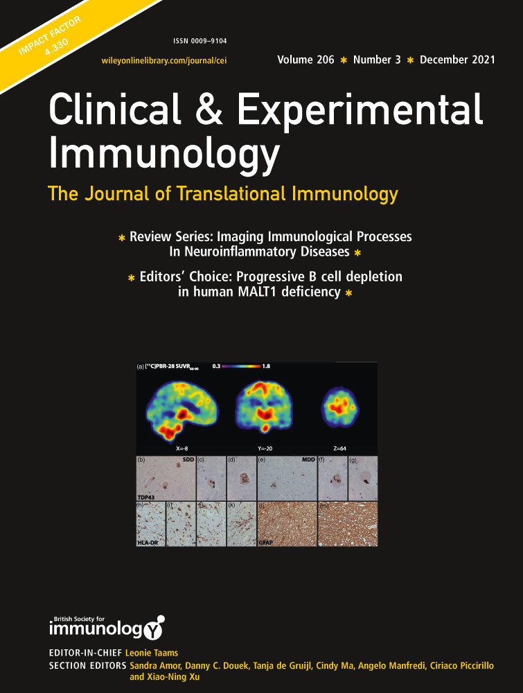
Imaging Immunological Processes from Blood to Brain in Amyotrophic Lateral Sclerosis
Neuropathology studies of amyotrophic lateral sclerosis (ALS) and animal models of ALS reveal a strong association between aberrant protein accumulation and motor neuron damage, as well as activated microglia and astrocytes. While the role of neuroinflammation in the pathology of ALS is unclear, imaging studies of the CNS support the idea that innate immune activation occurs early in disease in both humans, and in rodent models of ALS. In addition, emerging studies also reveal changes in monocytes, macrophages, and lymphocytes in peripheral blood as well as at the neuromuscular junction. To better understand the association of neuroinflammation (innate and adaptive) with disease progression the use of biomarkers and imaging modalities allow monitoring of immune parameters in the disease process. Such approaches are important for patient stratification, selection, and inclusion in clinical trials, as well as to provide readouts of response to therapy.
Here, we discuss the different imaging modalities e.g., MRI, MRS, PET as well as other approaches including biomarkers of inflammation in ALS, that aid the understanding of the underlying immune mechanisms associated with motor neuron degeneration in ALS.


留言 (0)