
記住我
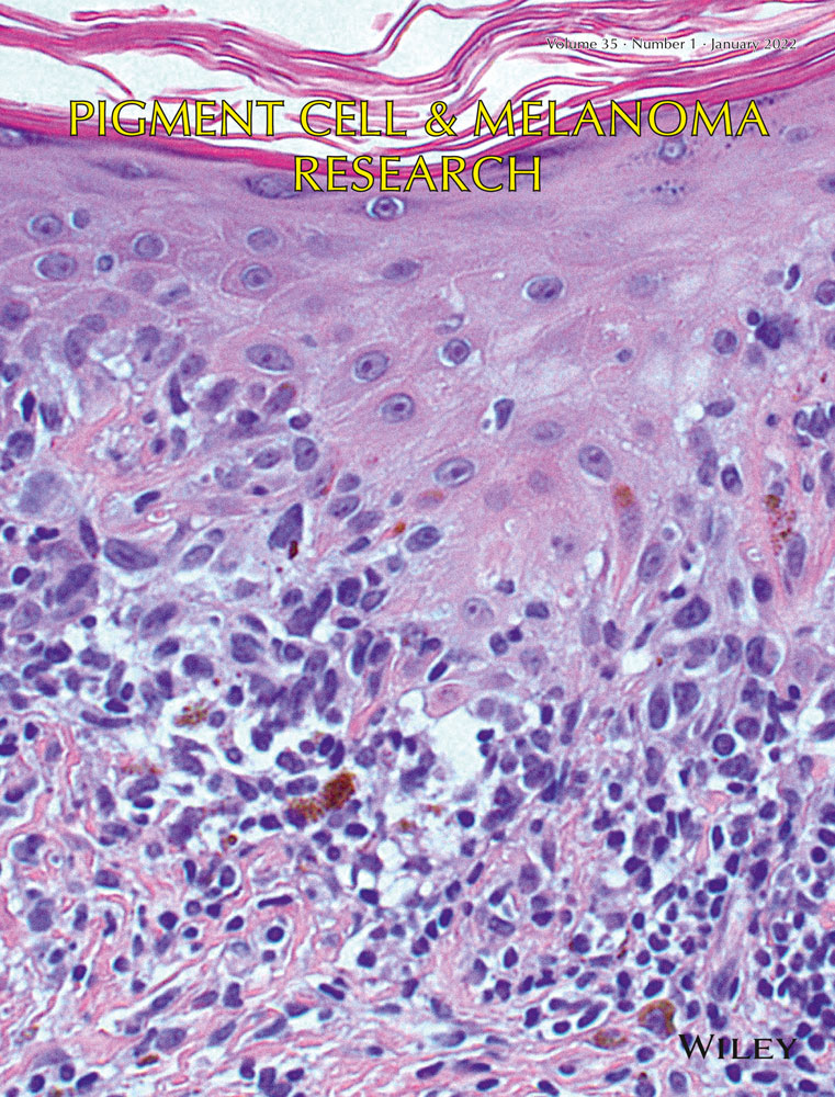


Melanoma is a heterogeneous disease that remains to be one of the most serious challenges for global health. Researchers have discovered several possible vaccination strategies for melanoma therapy. These strategies include vaccines targeting melanoma cells, peptide vaccines, DC-based vaccines, vector-based vaccines, and DNA vaccines (See Figure 1) (Rodríguez-Cerdeira et al. 2017).
The melanoma-associated antigen (MAGE) was the first tumor antigen (TA) identified for melanoma. Since its discovery, the MAGE gene family is targeted for vaccine design efforts against melanoma. Generally, TAs are classified into two main types including the mutational antigens and tumor-associated antigens (TAAs or self-antigens). The mutational antigens are not present in normal cells and are derived from mutated self-proteins. Some of these TA genes may be directly associated with the development of cancer including tumor suppressor and oncogenes genes such as Bcr-Abl, P53, and Ras. These antigens are also known as tumor-specific antigens (TSAs) and neoantigens (Lopes, Vandermeulen, et al., 2019). Tumor-associated antigens (TAAs or self-antigens) are the second class of TAs. These antigens do not harbor any mutations and are aberrantly expressed or overexpressed in cancer cells. TAAs include the products of silent genes, differentiation antigens, universal tumor antigens, and oncoviral TAAs. The silent genes encode for cancer/testis or oncofetal antigens such as melanoma-associated antigen (MAGE-1) and esophageal squamous cell carcinoma-1 (NY-ESO1) which are not expressed in postnatal tissues or normally expressed only in testis and placenta. The differentiation antigens such as gp100, tyrosinase, MART-1 (Melan-A), and TRP-1/- 2 are tissue-specific proteins that are overexpressed in tumor cells. The universal tumor antigens such as telomerase, survivin, and Her2/neu are expressed in low amounts in normal tissues but they are overexpressed in tumor cells. TAAs can include the oncoviral TAAs which are only expressed by malignant cells transformed after infection by an oncogenic virus such as Epstein–Barr virus (for nasopharyngeal carcinoma) and human papillomavirus (for cervical cancer) (Lopes, Vandermeulen, et al., 2019).
Vaccines ideally need to present all of these TAAs on the antigen-presenting cells (APC) to stimulate an adequate and long-lasting immune response. The posttranslational modifications, conformational changes, and gene overexpression could lead to the abnormal expression of TAAs in cancer patients including melanoma patients. These changes could overcome self-tolerance and stimulate the immune system to fight against cancer cells. The design of polyepitope TAA antigens provided the basis for the development of anticancer vaccines against multiple TAAs through the triggering of T-cell activation (Scardino, 2010). Vaccines that are designed against cancer antigens exert their antitumor effects by induction of cytotoxic T lymphocyte (CTL) response. These cells in turn recognize and attack the cancer-derived antigenic peptides presented by MHC molecules on the cancer cell surface. Different vaccination platforms including the use of protein, peptide, viral vector, mRNA, and DNA molecules can be used as vaccines (Marcela Valko-Rokytovská). DNA-based cancer vaccination strategy can be used to elicit immune responses against TAAs alone or in combination with antigens (multi-peptide) of cancer patients including melanoma (Scardino, 2010). The combination of the aforementioned vaccines was also studied in a “prime and boost” vaccination regimen to induce or extend the responses of the immune system. One of these prime/boost strategies is designed on the base of cancer antigens. Recently, DNA vaccines have been designed based on the different antigens of melanoma. This strategy successfully targets an antigen or multiple peptides of melanoma. For example, melanoma antigens including MART-1, tyrosinase, and melanoma-associated antigen 3 (MAGE-A3) are shown to be important in the induction of a balanced immune response. Therefore, they can be used individually or in a combinatorial strategy to design vaccines against melanoma cancer (Marcela Valko-Rokytovská).
3.1 Tumor-associated antigens (TAAs or self-antigens) 3.1.1 Tyrosinase antigenTyrosinase is a 75-kD copper-containing glycoprotein which is essential for pigmentation and melanin biosynthesis. It is a highly conserved protein present in many species including vertebrates, plants, and microbial organisms. Tyrosinase is known to induce immune responses against antigens related to melanogenesis in melanoma patients (Busam, 2019). Tyrosinase is stored in cytoplasmic organelles and synthesized by mucosal, epithelial, retinal, and ciliary body melanocytes. Melanin production is regulated by tyrosinase and tyrosinase-related protein 1 (TRP1). The melanin molecules can be metabolically derived from the oxidation of the L-tyrosine amino acid during a process catalyzed by tyrosinase. The enzymatic activity of tyrosinase is found in the sera of patients with metastatic melanoma which leads to the conversion of tyrosine to DOPA in stage II-IV melanosomes. Two membrane-bound enzymes with tyrosinase activity have been isolated from melanocytes. Both tyrosinase proteins are expressed in pigmented tissues. One of them is a membrane-bound tyrosinase and most of the tyrosinases in mammals belong to this type; the second type of tyrosinase is a soluble enzyme and exists to a lesser extent in the cells (Pragati Agarwal et al., 2019). Researchers have engineered a DNA vaccine based on mouse tyrosinase that has been administrated through the TriGrid delivery system. TriGrid delivery system is an investigational device that is dedicated to the clinical application. Compared to conventional injection methods, TriGrid can lead to enhanced intracellular delivery of vaccine constructs in skeletal muscle (Herrada et al., 2012). In another study, researchers demonstrated that intramuscular administration of the recombinant human and mouse tyrosinase could induce specific T-cell responses (Colluru et al., 2016).
Another strategy to improve the DNA vaccine efficacy is the simultaneous administration of tyrosinase epitopes (Synchrotope TA2M) into the groin lymph node. Synchrotope TA2M is a recombinant plasmid DNA vaccine encoding epitopes of tyrosinase with potential antineoplastic activities which is frequently expressed by melanoma cells. This strategy could lead to the production of anti-tyrosinase antibodies as well as the induction of CTL response against tyrosinase-expressing cancer cells. The elicited immune responses were shown to decrease tumor growth and improve survival in 16 out of 26 patients in clinical trials (Colluru et al., 2016). Several DNA vaccines based on the tyrosinase antigen are listed in Table 1.
Table 1. DNA vaccines based on the tyrosinase antigen Target antigen Tested model Adjuvant Device Site of administration Immune Response References Tyrosinase (xenogeneic/mouse) Human – TriGrid delivery system IM + EP Not describedYuan et al. (2013)
NCT00471133
Tyrosinase (human and mouse) Human – Biojector2000 jet delivery IM Specific T-cell responses Colluru et al. (2011) NCT00698100 Tyrosinase epitopes (Synchrotope TA2M) Human – Inguinal lymph node ID Induced immune responsesColluru et al. (2011)
NCT00023647
Tyrosinase (human) C57Bl/6 mice – – IM Inhibited MDSCs Yan et al. (2014) Tyrosinase (human) Dog – – – Induced antibody responses Manley et al. (2011) Tyrosinase (murine) Dog – Needle-free injection IM Induced antibody, T cell, and antitumor responses and increased survival Manley et al. (2011) Tyrosinase (human) Dog – Biojector2000 jet delivery IM Induced immune responses and long-term survival Wolchok et al. (2007) Tyrosinase (human) Dog Needle-free IM vaccination device TD Induced immune responses Colluru et al. (2011) Tyrosinase (canine) Cat – Vet Jet needle-free TD Induced immune responses with minimal risk of adverse effects Sarbu et al. (2017) Tyrosinase (human) Horse – Needle-free IM Antigen-specific immune responses Brown et al. (2013) 3.1.2 TRP1 (gp75) and TRP2 (DCT) antigensTyrosinase-related proteins (TYRPs) belong to a family of Zn2+/Cu2+ metalloenzymes sharing several sequence homologies. TYRPs are localized within specialized organelles called melanosomes. These proteins play crucial roles in a complex biochemical process known as melanogenesis and are consistently expressed in melanocytes of normal skin. The melanogenesis process leads to the formation of red pheomelanin and dark eumelanin. Two members of this family are known as Tyrp1 and Tyrp2. Both proteins have the same signal sequence, a transmembrane domain, two cysteine-rich domains, and two Zn2+/Cu2+ binding sites that are responsible for their catalytic activities (Ghanem & Fabrice, 2011).
The Tyrp1 (its mature form is originally called gp75) is encoded by the TYRP1 gene (the human homologue of the mouse brown locus) which is located in chromosome 9 (9p23). The gp75 is a 75-kDa transmembrane glycoprotein produced within the endoplasmic reticulum (ER) and is transported through the Golgi to melanosomes (Tie Fu Liu, 2001). According to published reports, Tyrp1 plays an important role in cell differentiation, oxidative stress, and activation of pigmentation. Tyrp1/gp75 is shown to be involved in melanocyte differentiation. Moreover, it is presumed to play an enzymatic role in the promotion of pigment formation. Abnormal synthesis of Tyrp1/gp75 and its interaction with calnexin (melanogenesis-associated chaperone) was shown to be responsible for the early cell death of vitiligo melanocytes due to their increased sensitivity to oxidative stress. TYRP1 mutations can also lead to a form of albinism in humans (oculocutaneous albinism type 3, OCA3). The tyrosinase protein expression in melanocytes does not vary with ethnicity. However, Tyrp1 protein was significantly elevated in Indian skin types and darkly pigmented Africans compared to lightly pigmented Chinese, Mexican and European skin types (Ghanem & Fabrice, 2011). The Tyrp1/gp75 has an important role in the activation of the pigmentation process through its enzymatic activity and regulation of tyrosinase activity. Although Tyrp1 implication in the melanogenesis process remains unclear, a series of biochemical experiments have demonstrated that Tyrp1/gp75 could act as catalase and dihydroxyindole carboxylic acid (DHICA) oxidase (Bertolotto, 2010). On the other hand, published reports exhibited that Tyrp1/gp75 may be contributed to the stabilization of tyrosinase. It has also been suggested that Tyrp1/gp75 and tyrosinase complex may prevent the premature death of melanocytes by attenuating tyrosinase-mediated cytotoxicity (Ghanem & Fabrice, 2011).
The Tyrp2 protein is encoded by dopachrome tautomerase (DCT) gene. This gene is located in chromosome 13 (13q32) with dopachrome and oxidoreductase activity which is involved in melanin and pigment color regulation. Diseases related to DCT include Vitiligo-Associated Multiple Autoimmune Disease Susceptibility (VAMAS) and Amelanotic Melanoma (Pak, 2006). The Tyrp2 expression was found in about 84% of primary and 60% of metastatic melanomas (Busam, 2019). The knowledge about the diagnostic potential of TRP1 and TRP2 is currently limited (Busam, 2019). There are very few studies on melanoma progression and TYRP1 expression, probably due to a melanocyte differentiation marker and its reputation as a melanogenic enzyme. Clinical studies have revealed that Tyrp1/gp75 could be the target in metastatic melanoma patients (Ghanem & Fabrice, 2011). In a study, the safety of injecting the gene (DNA) for mouse TYRP2 in patients with melanoma was investigated. Mouse TYRP2 DNA is very similar to human TYRP2 DNA. Based on laboratory experiments, it was determined that injection of mouse TYRP2 DNA could result in immune responses of T cells and antibody production. However, there is no still evidence that injection of mouse TYRP2 DNA results in any clinical benefit in patients (NCT00680589). Table 2 includes some DNA vaccine reports based on targeting TRP1 and TRP2 protein of the melanoma.
Table 2. DNA vaccines based on the TRP1 (gp75) and TRP2 antigens Target antigen Tested model Adjuvant Device Site of administration Immune Response References TRP1 Human – – IM Induced immune responsesGhanem and Fabrice (2011)
NCT00034554
TRP1 and TRP2 Mice – – – Induced immune responses Ghanem and Fabrice (2011) HLA-A*0201–restricted TRP1 epitope C57Bl/6 mice – Coated gold particles + gene gun provided by PowderMed IP Induced T-cell responses Ghanem and Fabrice (2011) TYRP2 (mouse) Human – – IM Not describedHerrada et al. (2012)
NCT00680589
TYRP2 + IgG1 C57Bl/6 mice – Coated gold particles Tail Induced T-cell responses Pudney et al. (2010) TYRP2 C57Bl/6 mice CCL21 HVJ anionic liposomes Leg Antigen-specific immune responses Yamano et al. (2006) TYRP2 C57Bl/6 mice CCL20, CCL21 EP Intratumoral or intradermal Antigen-specific immune responses Igoucheva et al. (2013) TYRP2 C57Bl/6 mice Hsp70 EP Oral Induced tumor-specific CTL response Zhu et al. (2010) TYRP2 C57Bl/6 mice Pattern recognitionreceptors (PRRs) EP ID Increased tumor-specific CTL response Lladser et al. (2011) TRP1 C57Bl/6 mice Alphaviral replicon Coated gold particles and gene gun IM or SC Induced innate antiviral pathways and improved responses Leitner et al. (2006) TRP1 C57Bl/6 mice Alphaviral replicase + Bcl-XL Gene gun SC Induced antibody responses Leitner et al. (2006) TRP1 and TRP2 C57Bl/6 mice IFN-gamma Gold complexes and helium-driven gun Intravenously via the tail vein Alternative roles Guevara-Patiño et al. (2019) 3.1.3 Gp100 antigenGp100 is a 100 kDa glycoprotein which is encoded by the PMEL (premelanosomal protein) gene. The GP100 is a structural component of the melanosomes and is a non-mutated differentiation antigen expressed on melanocytes that are overexpressed on melanomas. Modifications at positions 288 (valine for alanine) and 210 (methionine for threonine) produce peptides with increased binding affinity for HLA-A*0,201 MHC class I molecules (Busam, 2019). In a previous study, a DNA plasmid encoding for the modified gp100 was administered either intramuscular or intradermal to patients with metastatic melanoma. Then, the generation of anti-gp100 T cells and the impact on tumor proliferation were evaluated (Colluru et al., 2016). In the evaluation of mouse gp100 plasmid DNA vaccine, the vaccine was injected via IM or PMED in stage IIB, IIC, III, and IV melanoma patients. Patients are randomized to 1 of 2 treatment arms. Arm 1 of patients received vaccine by PMED on days 1, 3, 5, 8, 22, 24, 26, 29, 43, 45, 47, 50, 64, 66, 68, and 71. Arm 2 of patients received vaccine by IM injection on days 1, 3, 5, 8, 22, 24, 26, 29, 43, 45, 47, 50, 64, 66, 68, and 71. The elicited immune responses were shown to decrease tumor growth and improve survival in 4 out of 27 patients with increased CD8 + T cells and in 5 out of 27 patients with increased IFNγ + CD8+ responses in clinical trials (NCT00398073). Some of the studies based on the gp100 antigen have been listed in Table 3.
Table 3. DNA vaccines based on the gp100 antigen Target antigen Tested model Adjuvant Device Site of administration Immune response References gp100 (mouse) Human – Dermal PowderMed® IM or PMED Increased CD8 + IFN-γColluru et al. (2011)
NCT00398073
gp100 (mouse and human) Human – Biojector2000 jet delivery IM Specific T-cell responsesColluru et al., (2011)
NCT00104845
gp100 (human) Human – – IM or ID Antigen-specific immune responses Colluru et al. (2011) gp100 Human GM-CSF PowderJect XR1 PMED No enhancement in antigen-specific immune responses Colluru et al. (2011) gp100 Human – – IM or ID Inability to immunize in patients with metastatic Colluru et al. (2011) gp100 Human IL−2 – IM or ID Not described NCT00019448 gp100 C57Bl/6 mice MIP3α ECM 830 Electro Square Porator™ with 2-Needle Array™ BElectrode IM Induced immune responses and long-term survival Gordy et al. (2016) gp100 + CD40L C57Bl/6 mice GM-CSF and IL−2 – IM Enhanced survival and inhibited tumor growth Gupta et al. (2015) gp100 + Polysaccharides C57Bl/6 mice DsCE‑I Bio‑Rad Helios gene gun ID and SC Suppress tumor growth and prolong survival Wen-Chi Wei et al. (2014) gp100 C57Bl/6 mice – Water-in-oil-in-water SC Induced immune responses and inhibited tumor growth Kalariya and Amiji (2013) gp100 C57Bl/6 mice – HVJ-liposome IM Induced immune responses and long-term survival Yamano et al. (2006) gp100 C57Bl/6 mice IgG FC-CC21 Gene gun ID Enhanced immune responses Igoucheva et al. (2013) gp100 C57Bl/6 mice DC Liposome IP Induced immune responses Yang et al. (1999) 3.1.4 MART-1 (Melan-A) antigenMelanoma antigen (Melan-A) also known as melanoma antigen recognized by T cells (MART-1) is a structural component of the melanosome without enzymatic function. It is localized in the Golgi apparatus and outside the melanosome. Melan-A/MART-1 plays a central role in the expression, trafficking, stability, and processing of the melanocyte protein gp100. It is also involved in stage II melanosome biogenesis (Jacob Pitcovski et al., 2017) and usually expressed by intrafollicular and intraepidermal melanocytes in normal skin. The number of Melan-A/MART-positive melanocytes (60% to 90% of melanomas) is usually lower than TRP1 and tyrosinase-positive melanocytes but higher than GP100-positive melanocytes (Busam, 2019). Melan-A/MART-1 expression may be homogeneous in metastatic and primary melanomas. In particular, desmoplastic melanomas, such as spindle cell melanomas, are usually negative for Melan-A/MART1 (Busam, 2019). In a study, 12 patients with risk for relapse of melanoma were immunized with plasmid DNA encoding the MART-1 at doses of 0.1, 0.3, or 1.0 mg on days 1, 43, 85, and 127. Although systemic toxicity was not observed, also no enhancement in antigen-specific immune responses was seen (Colluru et al., 2016). Several MART-1 based antigen studies are listed in Table 4.
Table 4. DNA vaccines based on the MART-1 (Melan-A) antigen Target antigen Tested model Adjuvant Device Site of administration Immune Response References MART−1 + HBsAg Mice and human – – IM No enhancement in antigen-specific immune responses Colluru et al. (2011) MART−1 C57BL/6J mice DC Lipid lipoplexes IP Induced long-lasting immune responses Garu et al. (2016) MART−1 C57BL/6J mice DC Au-SGSH SC Induced a long-lasting immune responses KSuresh Kumar Gulla et al. (2019) MART−1 C57BL/6J mice – AuNPs-SL SC Induced a long-lasting immune responses Mokhtarzadeh, Parhiz, et al. (
留言 (0)