
記住我


E2A is expressed as two isoforms, E12 and E47, which are composed of approximately 600 amino acids. E2A contains five domains that are quite conserved in other E proteins (HEB and E2-2): ADs 1–3, the DES domain, and the bHLH domain. AD1 (amino acids 1–99) is located at the N-terminus of E2A. The DES domain (residues 100–239) is adjacent to AD1. AD3 (amino acids 211–298) and AD2 (amino acids 310–430) are in the middle of E2A. The bHLH domain (residues 547–608) is located at the C-terminus of E2A (Fig. 1) [10,11,12,13]. As transcriptional regulators, AD1, AD2, and AD3 of E2A and the NHR2-binding (N2B) motif mainly recruit transcriptional coregulators to initiate transcription, while the bHLH domain is mainly involved in the dimerization of E2A and binding to specific DNA motifs of target genes, leading to the execution of transcriptional regulation.
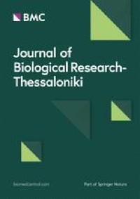
Fig. 1
Architecture of the domains of E2A. The domain architecture of CBP/P300 is shown in the middle, illustrating the four activation domains (AD1, DES, AD3, and AD2) and the C-terminal bHLH domain. The yellow shaded box represents AD1, and the core PCET motif of AD1 is represented by the small blue rectangle in the yellow shaded box. The dotted box represents the DES domain, and the N2B motif is represented by the small gold rectangle in the middle of the dotted box. The green box represents AD3, the orange box represents AD2, and the red box represents the bHLH domain. The numbers indicate the positions of the residues at both ends of these domains, AD1 (residues 1–99), DES domain (residues 100–239), AD3 (residues 211–298), AD2 (residues 310–430) and bHLH domain (residues 547–608). The structures of each domain are also shown and labeled. Top panel: The E2A PCET motif in complex with the ETO NHR1 domain (PDB: 2KNH); the E2A N2B motif in complex with the ETO NHR2 domain (PDB: 4JOL); the structure of the complex formed between the E2A bHLH domain and the SCL bHLH domain and E-box (PDB: 2YPB). Bottom panel: The PCET motif in complex with the CBP KIX domain (PDB: 2KWF); the structure of the complex formed between E2A AD1 (residues 1–37) and the CBP TAZ2 domain (PDB: 2MH0). The structures of the complex formed between E2A AD2 and NHR1, NHR2 or TAZ2 and the complex composed of E2A AD3 and TFIID was unclear
Among these ADs of E2A, AD1 is the most frequently studied and was the first AD discovered. In 1993, Aronheim et al. first identified AD1 in E2A when studying the mechanism underlying the transcriptional activation of target genes caused by E2A [11]. AD1 of human E2A is composed of 99 amino acids at the N-terminus. As one of the most well-conserved domains among the E proteins, E2A AD1 consists of 38 identical residues, 28 conserved residues and 33 relatively variable residues when compared with other E proteins (HEB and E2-2) (Fig. 2a). The three-dimensional (3D) structure of the full-length E2A AD1 has not been solved, and only NMR structures of the 37 N-terminal residues of AD1 (Met1–Pro37) have been reported [14]. The N-terminal region of AD1 forms a structure in which an α-helix and an adjacent short extended region are sandwiched by two coils. The first coil consists of 10 N-terminal residues (Met1–Val10). The 16 residues (Gly11–Phe26) adjacent to the first coil form an amphipathic α-helix. Pro27–Pro29 form a short extended region. The remaining eight amino acids (Val30–Pro37) comprise the second coil. The α-helix makes an important contribution to binding to the coactivator CBP or the corepressor ETO protein; therefore, this α-helix is called the P300/CBP and ETO target in the E protein (PCET) motif [15]. The stable structure of the α-helix and its adjacent short extended regions are important contributors to the interaction between proteins and AD1, and these regions are also conserved in other E proteins (Fig. 2b).
Fig. 2
The structure of AD1. a Alignment of AD1 in human E proteins (E2A, HEB, and E2-2). The blue shaded box denotes the core PCET motif, and the bottom symbols represent “*” identical residues, “:” conserved residues, and “.” semiconserved residues. The red dashed box represents the part of AD1 where the 3D structure has been reported. The LDFS motif is indicated by the red box. The amino acid sequences of human E proteins (E2A, HEB, and E2-2) were propagated from UniProtKB (P15923-2, Q99081-1, and P15884-1). b The 3D structure of a portion of E2A AD1 (residues 1–37). The 3D structure was derived from the Protein Data Bank (PDB: 2MH0); the blue helix denotes the PCET motif and is labeled; the two coils are shown in gray, and the short extended region is in blue
The PCET motif is the core element of AD1 and the most highly conserved region among the AD1 of E proteins [14, 16, 17]. As a transcriptional regulator, the transactivation function of AD1 is generally achieved by binding to transcriptional coactivators or corepressors, such as CBP or ETO [18,19,20]. The PCET motif (Gly11-Phe26) includes 7 hydrophobic residues (Leu16, Leu19, Leu20, Phe22, Met24, Met25, and Phe26), 4 amino acids with intermediate polarity (Gly11, Thr12, Ser17, and Ser23), 4 acidic residues (Asp13, Glu15, Asp18, and Asp21) and a basic residue (Lys14). The contribution of each residue of PCET to the interaction between PCET and CBP or ETO is slightly different. The distributed hydrophobic residues (Leu16, Leu19, Leu20, Phe22, Met24, and Met25) and the nearby hydrophilic residues constitute the core elements of PCET and are called the nine amino acid transactivation domain (9aaTAD) motif [21, 22]. The 9aaTAD plays a major role in the interaction between PCET and CBP or ETO and consists of two overlapping protein recognition motifs, Glu–Leu–Ser–Asp–Leu–Leu (ELSDLL) and Leu–Asp–Phe–Ser (LDFS). ELSDLL consists of three leucines (Leu16, Leu19, and Leu20) and the polar residues near these leucines. The ELSDLL sequence, which belongs to the general ɸ-X-X-ɸ-ɸ sequence, is present in the ADs of many transcription factors [18, 23, 24], where ɸ represents a bulky hydrophobic residue and X represents any amino acid residue. The four consecutive residues (residues 20–23) in the central portion of the 9aaTAD constitute the LDFS motif (Fig. 2a) [16, 25].
Interactions with CBP and ETOAs a transcriptional regulator, E2A recruits CBP or ETO to the promoter of acetylate or deacetylate histones, thereby leading to chromatin remodeling. The recruitment of CBP or ETO by E2A is mainly achieved through the interaction between the ADs of E2A and domains of CBP or ETO [26, 27]. AD1 generally interacts with the kinase-inducible domain interacting (KIX) domain and transcription adaptor putative zinc finger (TAZ2) domain of CBP and the TATA box-binding protein (TBP)-associated factor (TAF) homology (eTAFH, also called Nervy homology domain 1 (NHR1)) domain of ETO. The PCET motif is almost identical in E proteins. Based on the NMR structures of compounds containing AD1, namely, the E2A-AD1:TAZ2 complex, HEB-PCET:KIX complex, and HEB-PCET:eTAFH complex (Fig. 3) [14, 28, 29], the interactions between AD1 and these domains of CBP and ETO have many commonalities and some subtle differences.
Fig. 3
The interaction of AD1 with CBP or ETO. a Sharing mode of the interaction of AD1 with the domains of CBP or ETO. The residues Leu16, Leu19, and Leu20 of E2A are shown and labeled; the hydrophobic grooves on the surface of the domains of CBP or ETO are shown in gray and white. The 3D structure was obtained from the Protein Data Bank (PDB: 2MH0) and was modified using Discovery Studio 3.0 software. b Characteristics of the interaction of AD1 with NHR1/eTAFH. Top panel: The structure of eTAFH was obtained from the Protein Data Bank (PDB: 2KNH); the region marked in yellow on the domains represents the hydrophobic residues in the domains that bind to AD1; the polar residues on the domains that bind to AD1 are shown and labeled in red. Bottom panel: The sequences of HEB-PCET (12–27) were obtained from the Protein Data Bank (PDB: 2KNH); the hydrophobic residues in the sequences that contribute to the interaction with the NHR1/eTAFH domain of ETO are represented by yellow squares, and the polar residues involved in the interaction are marked in red. The red dotted line represents hydrogen bonds. c Top panel: The structure of KIX was obtained from the Protein Data Bank (PDB: 2KWF); the region marked in yellow on the domains represents the hydrophobic residues in the domains that bind to AD1; the polar residues in the domains that bind to AD1 are shown and labeled in red. Bottom panel: The sequences of HEB-PCET (12–27) are shown; the hydrophobic residues in the sequences that contribute to the interaction with the KIX domain of CBP are represented by yellow squares, and the polar residues involved in the interaction are marked in red. The red dotted line represents hydrogen bonds. d Top panel: The structure of the TAZ2 domain was obtained from the Protein Data Bank (PDB: 2MH0); the region marked in yellow on the domains represents the hydrophobic residues in the domains that bind to AD1; the polar residues of the domains that bind to AD1 are shown and labeled in red. Bottom panel: The sequences of E2A-PCET (11–29) were obtained from the Protein Data Bank (PDB: 2MH0); the hydrophobic residues in the sequences that contribute to the interaction with the KIX domain of CBP are represented by yellow squares, and the polar residues involved in the interaction are marked in red. The red dotted line represents hydrogen bonds
The commonalities are that the hydrophobic residues of the core PCET motif of AD1 insert into the hydrophobic groove on the surface of KIX, TAZ2 or NHR1; the hydrophobic residues of E2A interact with the amino acids in the hydrophobic groove through many nonpolar forces; and a small number of polar residues on PCET generate hydrogen bonds and electronic forces between residues of these domains to complement those nonpolar forces. More specifically, Leu16, Leu19 and Leu20 of PCET interact separately with residues in the three small hydrophobic pockets that comprise the hydrophobic groove (Fig. 3a), and the polar residue Glu15 interacts with polar residues outside the hydrophobic groove, namely, Asn627 of KIX, K273 of NHR1, and His1767 of TAZ2, through hydrogen bonds [14, 28, 29].
In addition, the interaction of AD1 with KIX, TAZ2 or NHR1 has its own characteristics. When PCET interacts with NHR1, perhaps due to a defect in the structure detection method, only Glu15, Leu16, Leu19 and Leu20 interact with the residues along the hydrophobic groove of NHR1 (Fig. 3b) [28]. When PCET interacts with KIX, in addition to Glu15, Leu16, Leu19 and Leu20 described above, the hydrophobic residues Phe22, Met24 (Ala25 in HEB), and Phe26 of PCET also interact with the residues in the hydrophobic cleft between helices H1 (Ile611, Phe612, Ala619, and Arg624) and H3 (Leu628, Tyr631, Ile660, Leu664, and Lys667) of KIX. The polar residue Asp21 of PCET interacts with Lys667 of KIX by forming a salt bridge, and Ser23 of PCET interacts with Thr614 of KIX by forming hydrogen bonds. A salt bridge also forms between Lys14 and Asp18 of E2A. Ser17 of PCET is exposed to solvent and does not contact any KIX residue. Gly11–Asp13 of PCET may have little contact with KIX due to flexibility (Fig. 3c) [29]. However, compared with the interaction between HEB-PCET and KIX, Leu20, Ser23, and Met24 are not involved in the interaction between E2A-AD1:TAZ2, while Asp18 and Met25 of PCET and Pro27 and Leu28 near the PCET motif are involved (Fig. 3d) [14]. In addition, the key residues of AD1 that bind to KIX, NHR1 or TAZ2 are also different. Leu20 in E2A is essential for E2A:KIX and E2A:eTAFH interactions [28, 29]. Leu19 and Phe22 in E2A are essential for E2A:TAZ2 interactions [14]. Key and major residues contributing to these protein interactions are located in the LDFS and 9aaTAD motifs. In addition, the three residues of the short extended region (Phe26, Pro27 and Leu28) contact the TAZ2 domain through extensive nonpolar contacts. Due to the insufficient details of the NMR structure of the known PCET-KIX complex and PCET-NHR1 complex, the role of the short extended region in the E2A:KIX and E2A:eTAFH interactions is unclear.
Although AD1 interacts with multiple domains on CBP and ETO, the roles of KIX, NHR1, TAZ2 and AD1 are generally independent and cooperative. The NHR1 binding site on AD1 is consistent with the KIX binding site, and the KIX domain of CBP and the NHR1 domain of ETO competitively bind AD1. Although both the KIX and TAZ2 domains of CBP bind to AD1, the key residues in their identified regions that bind to AD1 are slightly different; Leu20 is the key residue for the KIX interaction, whereas Leu19 and Phe22 are the key residues for the TAZ2 interaction [14, 28,29,30].
留言 (0)