Materials
Cytiva™ Ficoll-Paque™ PLUS Media and the Verso cDNA synthesis kit protocol were obtained from Thermo Fisher ScientificTM (Waltham, USA). INTEGRAGEN with NEBNext Ultra II DNA Library Prep Kit for Illumina was from New England BioLabs (Evry, FR). C18 SepPak cartridges was purchased from Waters (Guyancourt, FR). D-[2-3H]mannose (20 Ci/mmole) was obtained from Biotrends (Cologne, DE). En3hance spray and the kit chemagic STAR DNA Blood4k were purchased from PerkinElmer Life Sciences (Zaventem, BE). AG-1/AG50 columns, X-OMAT AR film and PNGase F from Elizabethkingia meningoseptica were obtained from SIGMA–Aldrich SARL (St Quentin Fallavier, FR). Thin Layer Chromatography (TLC) plates were purchased from MERCK (Darmstadt, DE). RNeasyⓇ Plus Mini Kit (50) was obtained from Qiagen (Courtaboeuf, FR). LightCyclerⓇ 480 SYBRⓇ Green I Master was purchased from Roche TM (Mannheim, DE).
Leukocyte isolation
Leukocytes were isolated from blood using a Ficoll-Paque Plus gradient. The genomic DNA was extracted from leukocytes employing the kit chemagic STAR DNA Blood4k according to the manufacturer’s instructions.
Cell culture
Skin biopsy fibroblasts from control and the patient, were cultured in RPMI medium supplemented with 10% FCS and 1% penicillin/streptomycin in 75 cm2 flasks, at 37 °C in a humid atmosphere containing 5% CO2.
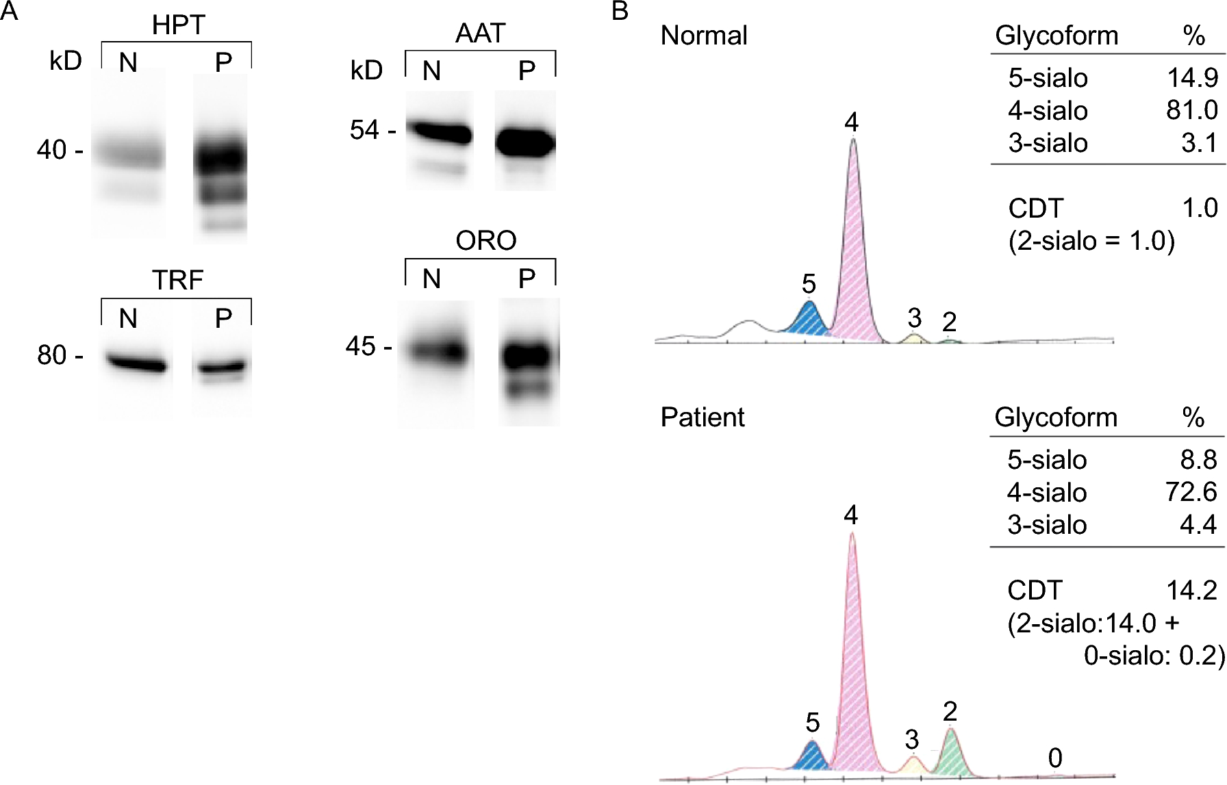
Western-blot of serum glycoproteins and enzyme activities
Western blotting of 4 different human serum glycoproteins (orosomucoid, alpha-antitrypsin, transferrin, and haptoglobin) were performed as previously described (5). Phosphomannomutase (6,7) and phosphomannose isomerase (8) activities were performed on leukocytes as previously described.
Capillary Zone Electrophoresis
Transferrin glycoforms were analyzed by capillary zone electrophoresis using the Capillarys Capiflex 2 automated system manufactured by Sebia (Lisses, France).
Mutation analysis
Next-generation sequencing (NGS) of 42 genes involved in CDG, was performed. Whole genome sequencing was performed by INTEGRAGEN with NEBNext Ultra II DNA Library Prep Kit according supplier recommendations, on an Illumina NovaSeq as Paired End 150 reads. The pathogenicity of the missense variant was evaluated with ACMG criteria: gnomAD frequency, in silico prediction and recommendations from the ClinGen SVI Splicing subgroup for interpretation of loss-of-function (LOF) variants. Functional data was necessary to prove a LOF ALG12 pathogenic variant.
Metabolic radiolabeling of cells
Fibroblasts established from skin biopsy from a control subject and from the patient, were pulse-radiolabelled as previously described (9). Briefly, cells layers were washed twice with PBS and once with labelling medium consisting of glucose-free RPMI 1640 supplemented with 1 mM Glc, 2% dialysed FCS and 2 mM fucose. The cells were then incubated for 30 min at 37 °C in 1 mL of labelling medium containing 100 µCi of [2-3H]mannose (20 Ci / mmol). The reaction was stopped by adding 12 mL PBS (4 °C).
Extraction of Dolichol-linked oligosaccharides (DLO)
DLO were isolated by the Folch extraction method as in (9). After removing the last PBS wash, cells were scrapped in the presence of 2 mL of extraction buffer containing 100 mM Tris HCl / 4 mM MgCl2 (pH 7.4) and methanol (1:2, v/v). The 2 ml cell extracts were collected in 15 ml tube on which the same volume of chloroform (CHCl3) was added. The tubes were then shaken vigorously, and after standing for 30 min at room temperature and centrifugation (room temperature, 1150 g, 5 min), the upper methanolic phase, the lower CHCl3 phase and the proteins at the interface, were recovered. After washing with methanol, 3 extractions of the protein interface were performed by adding a CHCl3/methanol/H2O mixture (10:10:3, v/v/v). Three 10/10/3 extractions were combined with the previously isolated CHCl3 phase to give a fraction containing total DLO.
Recovery of oligosaccharides from both radioactive DLO and glycoproteins
The CHCl3/10:10:3 samples were dried under vacuum and lipids were solubilized with 200 µL tetrahydrofuran. 2 mL of 22 mM HCl was added to the tubes and incubated at 100 °C for 45 min in order to release the oligosaccharides from the DLO. The glycoproteins from the 10:10:3 extracted protein pellet were submitted to Peptide N-glycanase digestion as described in the manufacturer’s instructions. Released N-glycans were desalted on AG-1/AG50 columns and further purified by passage through C18 SepPak cartridges. After drying under vacuum oligosaccharides were resolved by thin-layer chromatography (TLC) on silica-coated plastic sheets (0.2 mm thickness) that were developed in n-propanol/acetic acid/water, 3/3/2 for 20 h. Radioactive components were detected on X-OMAT AR film by fluorography after spraying the TLC with En3hance.
RT-qPCR ALG12
RNAs were extracted from control and patient fibroblasts according to the protocol from the RNeasyⓇ Plus Mini Kit (50) (QiagenTM). Retro-transcription (RT) was performed according to the Verso cDNA synthesis kit protocol (Thermo scientificTM) using 1 µg of RNA.
The cDNA sequence of ALG12 from exon 1 to exon 10 was amplified from 1 or 2 µl of the RT reaction by classic PCR using the primers F1 5’-TCGGGCTCTGTTCTGTTTCC-3’ and R1 5’-CCCTCGTTCTTTGGTGCTGA-3’ and further sequenced on both strands. Two couples of ALG12 primers were used for RT-QPCR: F2 5’-GCCAGTGGTGATCGCAGT-3’/R2 5’-CGAGTCCAAGCACTCCTCTAA-3’ and F3 5’-CCCATGCTCAACATCACG-3’/R3 5’- GCTTTGTACAGCCAAGACTTTTT-3’ for getting an amplicon partially covering respectively exon 3/exon 4 and exon 7/exon 8. QPCR was performed with the LightCyclerⓇ 480 SYBR Green I Master (RocheTM). In order to normalize the QPCR, the cDNA of the housekeeping gene ꞵ2M (ꞵ−2-microglobulin) was amplified with the F8 5’-GAGCATTCAGACTTGTCTTTCAGC-3’ / R8 5’-AATTCATCCAATCCAAATGCG-3’ primer couple. The ALG5 cDNA was amplified as an internal control with the F7 5’-TACACGTTGAACGATGGGCA-3’/R7 5’-TTCCGAGTTTGCTCAAGCCT-3’ primer couple.


留言 (0)