
記住我
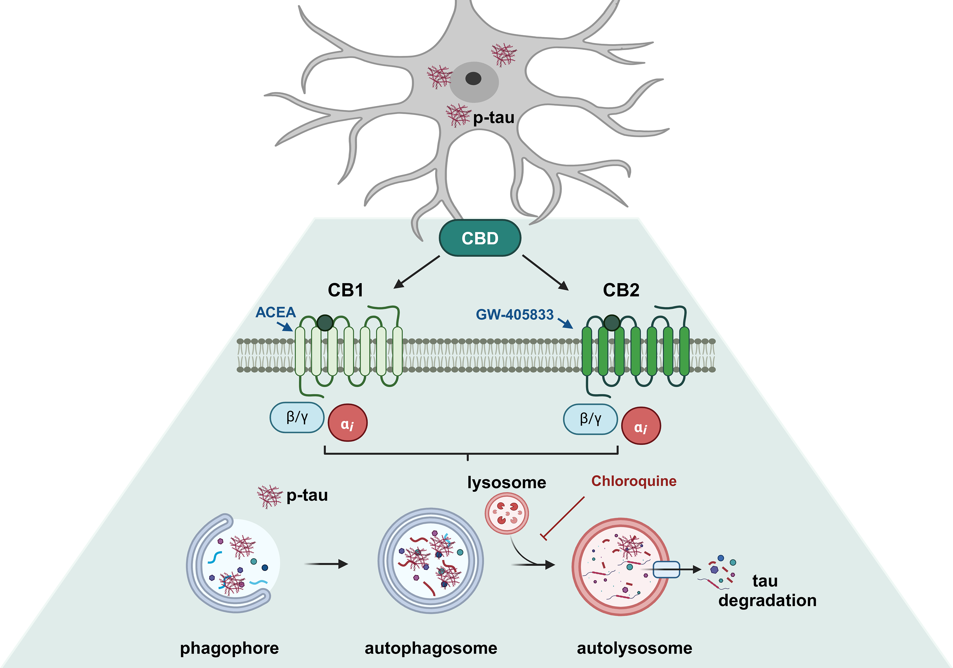


To characterize the overexpression of EGFP-Tau WT construct (~ 90 kD), the expression of total tau (Tau-5) was evaluated after 72 h of system activation with doxycycline (1 µg/mL) (Suppl. Figure 1 A-B). Data demonstrated the increase in tau-5 expression in the group treated with doxycycline, when compared to non-treated control group. Immunoblot analysis also showed no variation in endogenous tau when the Tet-On system is activated. After the activation of Tet-On system, the toxicity of cannabidiol and the selective agonists ACEA (CB1) and GW-405,833 (CB2) receptors, were evaluated. As shown in Fig. 1, there were no significant alterations in cell viability in the groups.
Fig. 1
Concentration-response curve of CBD, ACEA and GW-405,833 in the EGFP-Tau WT cell line. Cell viability was obtained from the colorimetric assay after Tet-On system activation with doxycycline (1 μg/mL) for 72 h and treatments with (A) CBD (100, 250 nM, 1 and 10 μM), (B) ACEA and GW-405,833 (100, 250 nM, 1 and 2 μM) for 24 h. Data are presented as viability of cell modulation in relation to the control without doxycycline. Data are presented as a mean ± SEM of five independent experiments (n=5; two-way ANOVA followed by Tukey’s post-test)
Cannabidiol Induces Tau ClearanceNext, after in Tau expressing SH-SY5Y cells (Dox+) presented a significant increase of tau-5 and its sites of phosphorylation AT8 (pSer202/pThr205) and AT180 (pThr231) (69% (p < 0.001); 50%, p = 0.0206 and 38.7%, p = 0.0490; respectively), when compared to the control (Dox−) (Fig. 2A-C). The results showed that the treatment with CBD significantly decreased the levels the phosphorylation sites at AT8 pSer202/pThr205 (tau AT8) by 35% (p = 0.020), 37% (p = 0.015) and 38% (p = 0.013), at 250 nM, 1 and 10 µM, respectively (Fig. 2A). On the other hand, no significant differences were observed in the AT180 phosphorylation (Fig. 2B). Additionally, CBD was able to decrease total tau (Tau-5) by 42% (p < 0.001), 64%, 64% and 50% (p < 0.0001) at 100, 250 nM, 1 and 10 µM, respectively, when compared to Dox+ (Fig. 2C). These data indicated that activation of Tet-On system induces the accumulation of tau total (Tau-5) and the phosphorylation of AT8 and AT180 proteins. Importantly, all concentrations tested of CBD abrogated the tau-5 and AT8 phosphorylation. Given this, we investigated the effect of CBD and the cannabinoid agonists ACEA and GW405833 on the AT8 phosphorylation site in the EGFP-tau-P301L cell line, which carries a mutated tau protein. Interestingly, CBD and ACEA at 1 µM after 24 h also reduced phosphorylation of AT8 in mutated tau by 30% (p = 0.0251) and 27% (p = 0.0465), respectively (Fig. 2D).
Fig. 2
CBD differentially affects the phosphorylation of AT8, AT180 proteins in EGFP-Tau WT and EGFP-Tau-PL301-expressing cell lines. EGFP-Tau WT cells were treated with CBD (100, 250 nM, 1 and 10 µM) for 24 h and cytosolic levels of AT8, AT180 and total tau (Tau-5) were evaluated by western blotting. EGFP-Tau-P301L cells were treated with CBD, ACEA and GW405833 at 1 µM for 24 h and cytosolic levels of AT8 were evaluated by western blotting. Data represent the mean obtained from the quantification of the optical density of the bands obtained by western blotting with the antibodies (A) anti-tau-5 (70-100 kD), (B) anti-pAT180 (70-100 kD), (C-D) anti-p-AT8 (70-100 kD) and normalized to the internal control α-tubulin (50 kD) or GAPDH (36 kD), as shown in the histograms (mean ± SEM). *p<0.05 and ****p<0.0001 vs. CTR (Dox–) and *p<0.05, ***p<0.001 and ****p<0.0001 vs. CTR (Dox+) (n=3-5; One-way ANOVA, followed by the Sidak post-test)
CBD Induces Autophagy in Tau WT Expressing SH-SY5Y Cell LineFurthermore, investigating the role of autophagy in tau degradation mediated by CBD. For this purpose, LC3-II protein was quantified in the EGFP-Tau WT cell line after treatment with CBD (100, 250 nM, 1 and 10 µM) for 2 h. The cells were also treated in presence and absence of ammonium chloride (NH4Cl, 2 mM), an inhibitor of the final step of autophagic flux, added in the last hour of treatment. As demonstrated in the representative immunoblots, the groups treated with CBD 100 nM and 10 µM had a further increase in LC3-II accumulation, 135.9% and 112.4% (p < 0.0001), when compared to NH4Cl (Fig. 3A). To evaluate the potential of CBD in inducing autophagy, the EGFP-Tau WT cell line overexpressing mCherry-LC3 was treated with CBD at 100 nM and 10 µM for 2 h, as previously demonstrated to activate autophagy. As a positive control, the cells were subjected to nutritional deprivation (starvation - STV) by Earle’s Balanced Salt Solution (EBSS), also for 2 h. The number of autophagosomes was performed in single-cells analysis under a confocal microscope (Fig. 3B). The data showed that there was an increase of mCherry puncta (LC3-II) of 96.1% (p < 0.0001), 95.4% (p = 0.0001) and 96.1% (p = 0.0097), in the CBD (100 nM), CBD (10 µM) and STV treated groups, respectively, when compared to the control (Dox+) (Fig. 3C). At the same concentrations, that CBD increased the levels of LC3-II protein in the immunoblot, it also induced the formation of autophagosome, which were observed in cells transfected with mCherry-LC3. In addition, to investigate the effect of CBD on autophagy in mutated tau, SH-SY5Y cells expressing EGFP-Tau-P301L were used. In contrast to prior findings, CBD and the selective cannabinoid agonists ACEA and GW-405,833 at 1 µM for 2 h did not modulate the levels of LC3-II (p = 0.9844) or p62 (p = 0.9749) (Fig. 3D-E, respectively).
Fig. 3
CBD and CB1/2 selective agonists differentially regulates autophagic flux in the EGFP-Tau WT or EGFP-Tau-PL301-expressing cell lines. (A) Cytosolic protein extracts were prepared and the autophagic flux was evaluated by western blotting in the EGFP-Tau WT cells treated with CBD (100, 250 nM, 1 and 10 µM) for 2 h in the presence or absence of NH4Cl (10 mM), added in the last hour of treatment. Mean LC3-II expression levels were compared between the CBD-treated groups, the control group (no treatment) and the NH4Cl group. Data represent the average optical density obtained by western blotting analysis with the anti-LC3-II antibody (15 kD) and normalized with the internal control for the anti-α-tubulin antibody (50 kD) as shown in the histograms (mean ± SEM). ****p<0.0001 vs. NH4Cl group (n=3; two-way ANOVA, followed by Tukey’s post-test). (B) Representative images of the EGFP-Tau WT cell line overexpressing the mCherry-LC3 plasmid and treated with CBD (100 nM and 10 μM) or submitted to nutritional deprivation (starvation– STV) for 2 h. (C) Quantification of mCherry-LC3 positive puncta (autophagosomes) after treatments with CBD (100 nM and 10 μM) and STV for 2 h. Images were obtained from at least 6 different fields, in a Carl Zeiss LSM 780 confocal microscope, at 63× magnification. Scale bar 10 μm. mCherry-LC3 Excitation/Emission: 543 / 615 nm Data are expressed as mean ± SEM **p<0.01, ***p<0.001 and ****p<0.0001 vs. CTR (One-way ANOVA, followed by Dunnett’s post-test). Cytosolic protein extracts were prepared and the autophagic flux was evaluated by western blotting in the EGFP-Tau-PL301 cells treated with CBD, ACEA and GW-405,833 (1 µM) for 2 h in the presence or absence of NH4Cl (10 mM), added in the last hour of treatment. (C) Mean LC3-II and (D) p62 expression levels were compared between CBD, ACEA and GW-405,833 treated groups, the control group (no treatment) and the NH4Cl group. Data represents the average optical density obtained by western blotting analysis with the anti-LC3-II antibody (15 kDa) or anti-p62 antibody (62 kDa) and normalized to the internal control GAPDH (36 kDa) as shown in the histograms. Data are expressed as mean ± SEM, vs. CTR or NH4Cl groups (n=3; Two-way ANOVA, followed by Tukey’s post-test)
CBD-Induced Autophagy Reduces Tau Accumulation Via AutophagyAs previously demonstrated, CBD induces autophagy and a tau reduction after 2 and 24 h of treatment. Using selective CBR agonists, EGFP-Tau WT-overexpressing cells were treated at different concentrations CBD, ACEA and GW405833 in association with the autophagic inhibitors: wortmannin (250 nM) and chloroquine (25 µM). Wortmannin is a specific inhibitor of phosphoinositidine 3-kinases (PI3Ks), used as an inhibitor of autophagy since it inhibits the initiation of the autophagosome (HUNG et al., 2009). Chloroquine is an autophagic blocker since it interferes in the fusion of autophagosome and lysosome. Thus, after the activation of Tet-On system, cells were pretreated with the autophagic inhibitors for 30 min, followed by CBD treatment and the selective agonists of CB1 and CB2, ACEA and GW405833 (1 and 2 µM), respectively for 24 h. The quantification of GFP fluorescence demonstrated that CBD (1 and 10 µM) decreased the fluorescence intensity of EGFP-Tau WT by 34.8% (p = 0.014) and 39.9% (p = 0.004), respectively, when compared to the control (Dox+) (Fig. 4A). The association of CBD with wortmannin did not affect the fluorescence intensity when compared to the control group. The groups co-treated with chloroquine increased 45% and 46% (p = 0.005) in fluorescence intensity in the groups treated with 1 µM and 10 µM CBD, respectively. These data demonstrated that chloroquine blocked the autophagic flux, causing the accumulation of tau and, consequently, increased the intensity of GFP fluorescence.
Fig. 4
EGFP-Tau fluorescence intensity levels modulated by CBD, ACEA and GW-405,833 in the presence and absence of autophagic inhibitors. Cells were activated with doxycycline (1 µg/mL) for a period of 72 h, pre-treated with wortmannin (250 nM) and chloroquine (25 µM) for 30 min and then treated with CBD (100, 250 nM, 1 and 10 µM), ACEA (100, 250 nM, 1 and 2 µM) and GW-405,833 (100, 250 nM, 1 and 2 µM) for 24 h. Representative images for CBD (1 and 10 µM), ACEA (1 and 2 µM) and GW-405,833 (1 and 2 µM) were shown. (A) Data represent the means of tau fluorescence intensity after treatment with CBD, obtained by the analysis and normalized with the control group (mean ± SEM) *p<0.05 and **p<0.01 vs. CTR group; @@p<0.01 vs. respective group without inhibitors (n=7; two-way ANOVA, followed by the Sidak post-test). (B) Data represent the means of tau fluorescence intensity after treatment with ACEA obtained by the analysis and normalized with the control group (mean ± SEM). *p<0.05 and **p<0.01 vs. CTR group; @p<0.05 and @@p<0.01 vs. respective group without inhibitors (n=7; two-way ANOVA, followed by the Sidak post-test). (C) Data represent the means of tau fluorescence intensity after treatment with GW-405,833 obtained by the analysis and normalized with the control group (mean ± SEM). *p<0.05, **p<0.01 and ***p<0.001 vs. CTR group; @p<0.05 and @@p<0.01 vs. respective group without inhibitors (n=7; two-way ANOVA, followed by the Sidak post-test)
Cells treated with ACEA showed a significant decrease of 54.6% and 42.9% (p = 0.001) in the fluorescence intensity of EGFP-Tau WT at 1 and 2 µM, respectively (Fig. 4B). The association of ACEA and wortmannin caused a decrease of 23.9% (p = 0.004) of EGFP-Tau WT fluorescence when compared to control. Additionally, wortmannin at 1 and 2 µM led to a decrease of 41% (p = 0.027) and 40% (p = 0.025) in fluorescence intensity in relation to the control, respectively.
The co-treatment with chloroquine decreased in 47.7% (p = 0.017) and 49.5% (p = 0.005) compared to the control and 85.2% (p = 0.017) and 58.6% (p = 0.010) at 100 and 250 nM, respectively.
Cells treated with GW-405,833 showed a significant decrease of 59.3% and 69.9% (p = 0.001) in the EGFP-Tau WT fluorescence intensity at 1 and 2 µM, respectively (Fig. 4C). Co-treatment of GW-405,803 with wortmannin led to a further decreased of 51% (p = 0.001) e 51.1% (p = 0.0.007) in fluorescence intensity in relation to the wortmannin control, at 100 nM and 1 µM, respectively. Also, GW-405,803 was able to decrease 24.8% (p = 0.002) and 75.8% (p = 0.027) in relation to their respective groups without inhibitors, control group and group treated with 100 nM, respectively. The co-treatment with chloroquine showed a reduction of 47% (p = 0.010) in relation to chloroquine alone and an increase of 93.03% (p = 0.034) in the group treated with 2 µM in relation to its non-treated group (Fig. 4C). In summary, these data showed that the cannabinoids CBD, ACEA and GW-405,833 decreased the intensity of tau fluorescence. However, when using the autophagic inhibitors, wortimanin and chloroquine, only the CBD (1 and 10 µM) and GW-405,833 (2 µM) + chloroquine groups, reversed the decrease in the intensity of EGFP-Tau WT, demonstrating the possible participation of autophagy in the degradation of tau in these groups.
留言 (0)