NASCI case of the month: ‘Intramyocardial dissecting hematoma’
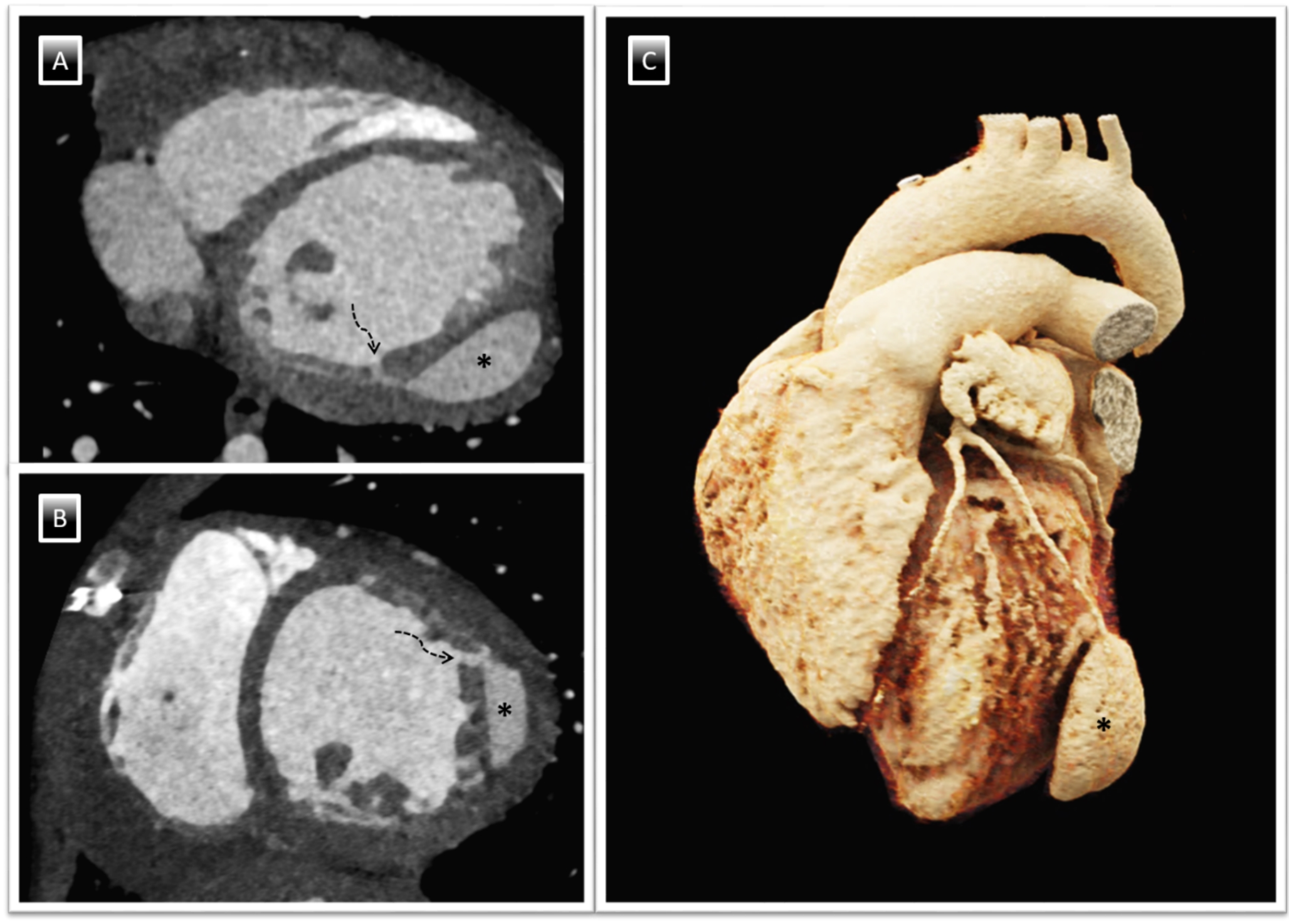
Intramyocardial dissecting hematoma (IDH) usually develops from hypoxia-induced capillary destruction within the myocardium following ischemia. The hematoma then infiltrates the interstices between myocardial spiral fibers, establishing a neocavity. As intra-neocavitary pressure increases, the hematoma expands and occasionally becomes associated with the epicardium or ventricular cavity (Roslan A et al. (2017) Intramyocardial dissecting hematoma in patients with ischemic cardiomyopathy: role of multimodality imaging in three patients treated conservatively. CASE: Cardiovasc Imaging Case Rep 1(4):159). Differential diagnoses include prominent ventricular trabeculations, intracavitary thrombosis, and pseudoaneurysm. By confirming the integrity of the epicardium, IDH can be distinguished from pseudoaneurysm, characterized by a complete rupture of the myocardial wall enclosed by the pericardium. Clear identification of the endocardium surrounding the hematoma and its systolic expansion may help to differentiate IDH from intracavitary hematoma. Prominent trabeculations can be recognized by a ventricular wall with an utterly asymmetric flow pattern (Vasco Dias (June 2009) et al., Intramyocardial dissecting haematoma: a rare complication of acute myocardial infarction. Eur J Echocardiography 10(4):585–587).


留言 (0)