Human placental tissue collection and isolation of primary human trophoblast cell
All placentas were voluntarily donated by patients at the First Affiliated Hospital of Chongqing Medical University with informed consent and institutional approval. The Human Research Ethics Board of the Chongqing Medical University approved the human placental tissues collection procedures (#2,022,127). Placental tissues were collected from normal pregnant women with voluntary termination of pregnancy (n = 3, gestational age 6–8 weeks), those with PE (n = 6, gestational age 36–38 weeks), and age-matched normal controls (n = 6, gestational age 38–40 weeks). The diagnostic criteria for PE patients refer to the American College of Obstetrics and Gynecology (ACOG) guideline that new-onset hypertension (systolic blood pressure ≥ 140 mmHg and/or diastolic blood pressure ≥ 90 mmHg; average of two measurements) after 20 weeks of gestation with proteinuria or other organ damage [15]. The exclusion criteria included women with other systemic diseases, such as preexisting hypertension, cardiovascular diseases, renal disease, immune disease, or other gestational complications. To isolate primary human trophoblast cells (PHT), normal term placenta was washed twice with normal saline, cut into small pieces using sterile scissors, and then digested with an enzyme mixture containing trypsin (Sigma-Aldrich, St. Louis, MO, USA), DNase (Roche, Basel, Switzerland), and Dispase (Sigma-Aldrich). Percoll (GE Healthcare, Chicago, IL, USA) gradient centrifugation was utilized to enrich VCT, and the collected cells were cultured in DMEM medium (Sigma-Aldrich) supplemented with 10% fetal bovine serum (FBS, Gibco, Waltham, MA, USA). VCT cells spontaneously fused into SCT cells in 48 h of culture in vitro, so VCT cells and SCT cells were collected separately after 3 h and 48 h of culture.
Cell culture
BeWo cells (American Type Culture Collection, ATCC, Manassas, VA, USA) were cultured in F12K medium (ATCC) supplemented with 10% FBS. To induce cell fusion, BeWo cells were treated with 25 µM forskolin (FSK, Sigma-Aldrich) for 48–72 h. HEK-293T (Cell Bank, Chinese Academy of Sciences) was cultured in DMEM (Gibco) supplemented with 10% FBS. All cells were cultured in 37 °C, 5% CO2 incubator. Prior to use, all cell lines were tested negative for mycoplasma contamination using a mycoplasma detection kit (TransGen Biotech, Beijing, China).
Mass spectrometry
Human placental villi from early pregnancy were collected and then lysed on ice for 20 min in 1 ml of RIPA buffer (Beyotime, Shanghai, China) supplemented with 1 × Phenylmethanesulfonyl fluoride (PMSF, Beyotime). Whole cell lysates were subjected to immunoprecipitation (IP) overnight at 4 °C using P57 antibody (Cell Signaling Technology, Danvers, MA, USA; catalog: #2557S) or normal rabbit IgG (Cell Signaling Technology, Danvers, MA, USA; catalog: #2729). The 25 µl prewashed Protein A/G magnetic beads (Thermo Scientific Pierce, Waltham, MA, USA; catalog: 88,802) were added to the lysis buffer and antibody mixture and incubated at room temperature for one hour. IP protein was separated by SDT buffer (4% SDS, 100 mM Tris-HCL, 1 mM DTT, PH = 7.6). Peptide detection was performed by liquid chromatography-tandem mass spectrometry (LC-MS/MS) in Applied Protein Technology (Shanghai, China), and coupling analysis was performed with Easy NLC by Q Exactive mass spectrometer.
Plasmid construction
The plasmids utilized in this study were generated through following steps: the eukaryotic expression vector pCDH was first linearized by restriction endonuclease, ECoR-I (New England Biolabs, NEB, Beverly, MA, USA) and Not-I (NEB), and the linearized carriers were then recovered by agarose gel electrophoresis and using a gel recovery kit (Omega Bio-Tek, Norcross, GA, USA). Fragments comprising STK40 (Genbank id: NM_001282547.2), P57 (Genbank id NM_001122630.2), and ubiquitin (UB; Genbank id: X04803.2) were obtained from the human placental villus cDNA library and fused with HA or MYC tag, then cloned into pCDH vector using homologous recombination, respectively. All fragments were generated using high-fidelity polymerase (TransGen Biotech, China) and homologous recombination was performed by Exnase II ligase (Vazyme, Nanjing, China). Finally, the STK40 vector containing the HA tag, P57 truncated mutant containing MYC tag, and HA-tag Ub vector were obtained. The COP1 vector was purchased from Gene Copoeia (Guangzhou, China; Genbank id: NM_022457.7). Synthesized shRNA targeting STK40 (Genbank id: NM_032017.3, 5′-CCGGATGGTTAAGAAGATGAA-3′) was cloned into a pSIH1 vector to construct knockdown of STK40 vector.
Lentivirus packaging and infection
HEK-293T cells were cultured in 60 mm dishes until reaching 60–80% confluence. A mixture of psPAX2, pVSVG, the target plasmid, and polyethylenimine Linear transduction reagent (Polysciences Co., USA) was added to the cells. Lentiviral particles were collected 48–72 h after inoculation. The collected virus particles were used to infect BeWo cells after adding 2 µg/ml polybrene. After 12 h of infection, cells infected with pCDH or shRNA were selected with 6 µg/ml puromycin. The selected cells were re-inoculated into a six-well plate containing F12K complete medium supplemented with 1 µg/ml puromycin and 10% FBS to continue culture to obtain stable overexpression or knockdown of STK40 cell lines.
Co-immunoprecipitation (Co-IP)
To extract protein, samples of human villus tissue or cells were lysed in a buffer containing protease and phosphatase inhibitors. The resulting mixture was centrifuged at 12,000 g for 15 min at 4 °C to obtain the supernatant containing the protein. Additionally, a tenth of the lysate was extracted as an input sample. The remaining lysates were divided into two equal parts. Subsequently, the target antibody and IgG were separately added to each part, followed by overnight incubation at 4 °C. Afterward, 25 µl prewashed Protein A/G magnetic beads were introduced to the lysate and antibody mixture, and the solution was incubated at room temperature for one hour. Proteins associated with Protein A/G beads were collected using a magnetic rack. The protein was eluted with SDS polyacrylamide gel electrophoresis (SDS-PAGE) washing buffer and subjected to Western blot (WB).
Immunofluorescence
Cells were seeded on a sterile glass slide and placed in a 24-well plate with or without FSK treatment. After incubation, the cells were gently washed three times with phosphate buffered saline (PBS), and fixed with 4% paraformaldehyde for 15 min. Following fixation, the cells underwent three additional washes with PBS and were permeabilized with a 0.1% Triton X-100 PBS solution for 15 min. To minimize non-specific binding, the cells were incubated in a PBS solution containing 3% bovine serum albumin (BSA) for an hour. Next, the primary antibody was added to the cells and allowed to incubate overnight at 4 °C. Details of antibodies were as follows: Vimentin (1:200, Bioss, Woburn, MA, USA; catalog: BS-0756R), Cytokeratin-7 (CK7, ZSbio, Beijing, China; catalog: ZM-007), STK40 (1:200, Biorbyt, Cambridge, United Kingdom; catalog: orb101780), E-Cadherin (E-CAD, 1:200, CST; catalog: #14,472). After three washes with PBS, the cells were stained with the corresponding goat anti-rabbit IgG (H + L) cross-adsorbed secondary antibody Alexa Fluor 488/goat anti-mouse IgG (H + L) cross-adsorbed secondary antibody Alexa Fluor 594 (1:1000; Thermo Fisher Scientific) and incubated for an hour. Finally, the cells were counterstained with DAPI (1:1000; Beyotime) to visualize the nucleus. Immunofluorescence (IF) signals were captured by using confocal laser scanning microscopy (CLSM) (Nikon, Tokyo, Japan).
Protein extraction and western blot
Whole cells were lysed in RIPA buffer (Beyotime) supplemented with 1 × PMSF, and the protein concentration was determined using the BCA protein assay kit (Beyotime). All protein imprinting was performed using a micro-electrophoresis system (Bio-Rad, Hercules, CA, USA). The total protein was separated on a 10% SDS-PAGE gel and transferred to a polyvinylidene fluoride (PVDF) membrane (Millipore, Burlington, MA, USA). Following blocking with 5% skim milk at room temperature for an hour, the membrane was incubated with primary antibodies overnight at 4 °C with the following primary antibodies: STK40 (1:1000, Invitrogen; catalog: PA5-22165), P57 (1:1000, Proteintech, Wuhan, China; catalog: 66794-1-Ig), CGβ (1:1000, Proteintech; catalog: 11615-1-AP), E-CAD (1:1000, CST; catalog: #14,472), GAPDH (1:1000, Beyotime; catalog: AF0006), Myc-Tag (1:1000, CST; catalog: #2276), COP1 (1:1000, Abcam, Cambridge, MA, USA; catalog: ab56400), Ubiquitin (1:1000, Abmart, Shanghai, China; catalog: T55965). Subsequently, the membrane was incubated with the corresponding Goat anti-Rabbit/Mouse IgG (H + L) Secondary Antibody, HRP (1:10000, Thermo Scientific Pierce, C31460100/C31430100) at 37 °C for one hour. The target protein was detected using the ECL-Plus kit (NCM Biotech, Suzhou, China).
RNA isolation, cDNA synthesis, and RT-qPCR
Total RNA was isolated using Trizol reagent (Takara, Shiga, Japan). RNA concentration was measured by spectrometer (Bio-rad). For the synthesis of cDNA, 0.1–0.5 µg of total RNA was used for reverse transcription according to the reverse transcription kit (TransGen Biotech, China). real-time quantitative PCR (RT-qPCR) was performed using SYBR Green qPCR Master Mix (TransGen Biotech, China) and real-time PCR system (Bio-Rad). Primer sequences used were as follows: STK40 forward, 5′-CAGCACTACGTCATCAAGGAG-3′, STK40 reverse, 5′-CGATGTGTCCTCTCTTGTTGAGC-3′; P57 forward, 5′-GCGGCGATCAAGAAGCTGT-3′, P57 reverse, 5′-GCTTGGCGAAGAAATCGGAGA-3′; and GAPDH forward, 5′-GGAGCGAGATCCCTCCAAAAT-3′, GAPDH reverse, 5′-GGCTGTTGTCATACTTCTCATGG-3′. GAPDH was used as a standardized internal control. The specificity of the fluorescence signal was verified by melting curve analysis and gel electrophoresis. The expression level of the target gene was determined by the 2−ΔΔCT method.
Cell viability and proliferation assays
Cell viability was measured by Cell Counting Kit 8 (CCK8, NCM Biotech). Cells were seeded into 96-well tissue culture plates at a density of 3 × 10³ cells per well. At each time point, 10 µl of CCK8 solution was added to each well, and the plates were incubated at 37 °C for two hours. The absorbance at 450 nm was measured using a microplate reader (Molecular Devices, San Jose, CA, USA). Cell proliferation was determined using 5-ethynyl-2′-deoxyuridine (EdU) assay. Briefly, cells were inoculated in cell crawls at a density of 1 × 10⁴ cells and cultured overnight. The cells were then treated with 10 µM 5-Fu for 24 h. Next, 10 µM EdU (Beyotime) was added to the medium and incubated for an additional 12–24 h. Cells were fixed and stained according to the manufacturer’s instructions.
Cell cycle assay
STK40-OE and sh-STK40 cells and their control cells were seeded in a six-well culture plate and cultured to 80% confluence. Cell cycle analysis was conducted via nuclear Propidium Iodide (PI, Beyotime) staining, and PI absorbance was measured by BD FACSCelesta Multicolor Flow Cytometer (BD Biosciences, San Jose, CA, USA).
Rigid molecular simulation docking
The PDB format files of the 3D crystal structures of P57 and STK40 proteins were obtained from the UniProt protein database (https://www.uniprot.org/). The model calculation was carried out using the GRAMM-X protein-protein docking server (https://gramm.compbio.ku.edu/), with P57 was selected as the receptor and STK40 as the ligand to analyze the two full-length protein groups. Docked complexes were analyzed using the PDBePISA server (https://www.ebi.ac.uk/msd-srv/prot_int/pistart.html). Results are visualized using PyMOL (Version 2.5.7).
RNA sequencing
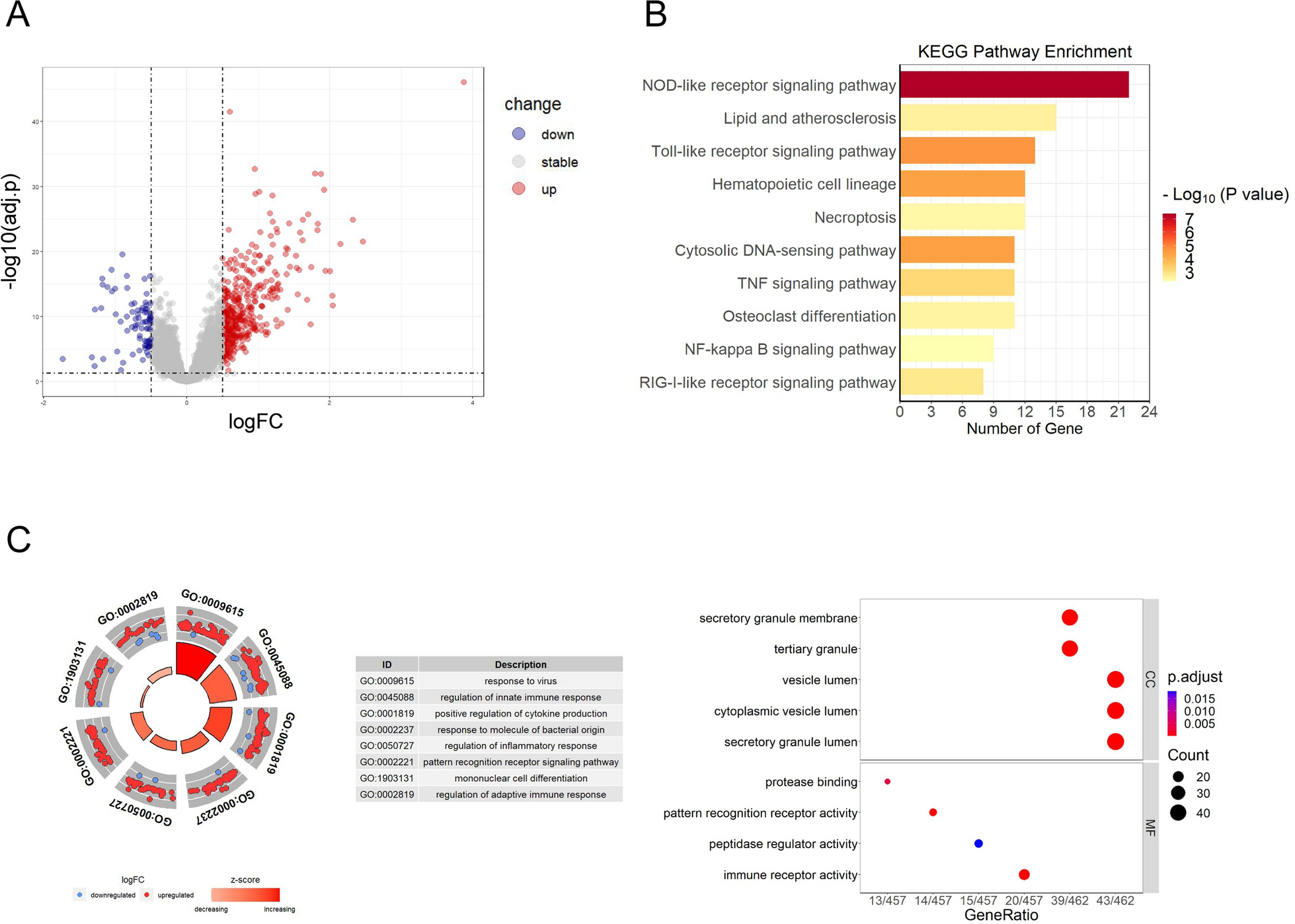
Stably transfected cell lines, including STK40-OE and sh-STK40, as well as their respective control counterparts, were harvested, and total RNA was extracted using Trizol (Takara). RNA integrity was evaluated through 1.0% agarose gel electrophoresis. Subsequently, the RNA that meets the required standards was sent to Personalbio (Shanghai, China) for cDNA library preparation and RNA-seq. Samples were indexed and sequenced on an Illumina NovaSeq 6000. Raw reads were quality-controled and cleaned using FastQC (Version 0.11.9) and fastp (Version 0.23.1), and then aligned to the human genome GRCh38.p13 using HISAT2 (version 2.2.1). Gene counts were derived using Subread: featureCount (version 2.0.3). The DESeq2 package in R (Version 4.3.1) was used to analyze differentially expressed genes (DEGs) in RNA-seq data. Genes with a p-value < 0.05 and an absolute fold change > 0.5 were considered DEGs. Volcano plots show the expression patterns of these DEGs. Gene ontology (GO) and Kyoto Encyclopedia of Genes and Genomes (KEGG) pathway analysis were used to explore the possible functions of these DEGs.
Statistical analysis
Statistical analysis was performed using GraphPad Prism software (Version 5.0). Each experiment was repeated independently at least three times. Significant changes between the two groups were analyzed by Student’s t-test. A value of P < 0.05 was considered significant, denoted as * P < 0.05, ** P < 0.01, and *** P < 0.001. All data are expressed as mean ± standard deviation (SD).


留言 (0)